Suppr超能文献

基于机器学习和基于颜色的图像集聚类的交互式引导图像分割。

Guided interactive image segmentation using machine learning and color-based image set clustering.

机构信息

Institute of Computer Science, Leipzig University, Leipzig 04107, Germany.

IfADo-Leibniz Research Centre for Working Environment and Human Factors, Dortmund 44139, Germany.

出版信息

Bioinformatics. 2022 Sep 30;38(19):4622-4628. doi: 10.1093/bioinformatics/btac547.

Abstract

MOTIVATION

Over the last decades, image processing and analysis have become one of the key technologies in systems biology and medicine. The quantification of anatomical structures and dynamic processes in living systems is essential for understanding the complex underlying mechanisms and allows, i.e. the construction of spatio-temporal models that illuminate the interplay between architecture and function. Recently, deep learning significantly improved the performance of traditional image analysis in cases where imaging techniques provide large amounts of data. However, if only a few images are available or qualified annotations are expensive to produce, the applicability of deep learning is still limited.

RESULTS

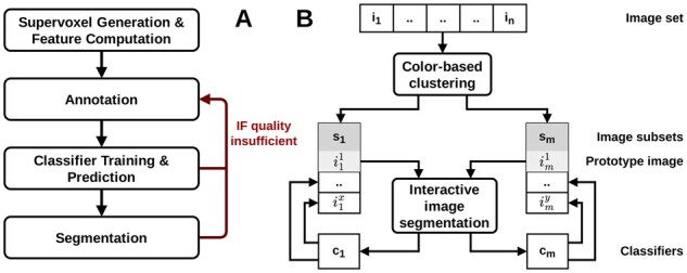
We present a novel approach that combines machine learning-based interactive image segmentation using supervoxels with a clustering method for the automated identification of similarly colored images in large image sets which enables a guided reuse of interactively trained classifiers. Our approach solves the problem of deteriorated segmentation and quantification accuracy when reusing trained classifiers which is due to significant color variability prevalent and often unavoidable in biological and medical images. This increase in efficiency improves the suitability of interactive segmentation for larger image sets, enabling efficient quantification or the rapid generation of training data for deep learning with minimal effort. The presented methods are applicable for almost any image type and represent a useful tool for image analysis tasks in general.

AVAILABILITY AND IMPLEMENTATION

The presented methods are implemented in our image processing software TiQuant which is freely available at tiquant.hoehme.com.

SUPPLEMENTARY INFORMATION

Supplementary data are available at Bioinformatics online.

摘要

动机

在过去的几十年中,图像处理和分析已成为系统生物学和医学中的关键技术之一。对活体系统中解剖结构和动态过程的定量分析对于理解复杂的潜在机制至关重要,并且允许构建阐明结构与功能之间相互作用的时空模型。最近,深度学习在成像技术提供大量数据的情况下,极大地提高了传统图像分析的性能。但是,如果可用的图像很少,或者生成合格的注释很昂贵,则深度学习的适用性仍然有限。

结果

我们提出了一种新方法,该方法结合了基于机器学习的超像素交互式图像分割和聚类方法,用于自动识别大型图像集中颜色相似的图像,从而可以引导重用交互式训练的分类器。我们的方法解决了由于生物和医学图像中普遍存在且通常不可避免的显着颜色变化而导致重新使用训练有素的分类器时分割和量化精度降低的问题。这种效率的提高提高了交互式分割对于更大图像集的适用性,能够以最小的工作量对深度学习进行高效的定量或快速生成训练数据。所提出的方法几乎适用于任何图像类型,并且是一般图像分析任务的有用工具。

可用性和实现

所提出的方法已在我们的图像处理软件 TiQuant 中实现,该软件可在 tiquant.hoehme.com 上免费获得。

补充信息

补充数据可在“生物信息学”在线获得。

https://cdn.ncbi.nlm.nih.gov/pmc/blobs/199c/9525009/586880203972/btac547f1.jpg

相似文献

1
Guided interactive image segmentation using machine learning and color-based image set clustering.
Bioinformatics. 2022 Sep 30;38(19):4622-4628. doi: 10.1093/bioinformatics/btac547.
2
TiQuant: software for tissue analysis, quantification and surface reconstruction.
Bioinformatics. 2015 Oct 1;31(19):3234-6. doi: 10.1093/bioinformatics/btv346. Epub 2015 Jun 3.
3
Trainable Weka Segmentation: a machine learning tool for microscopy pixel classification.
Bioinformatics. 2017 Aug 1;33(15):2424-2426. doi: 10.1093/bioinformatics/btx180.
4
PyMIC: A deep learning toolkit for annotation-efficient medical image segmentation.
Comput Methods Programs Biomed. 2023 Apr;231:107398. doi: 10.1016/j.cmpb.2023.107398. Epub 2023 Feb 7.
5
A review of image analysis and machine learning techniques for automated cervical cancer screening from pap-smear images.
Comput Methods Programs Biomed. 2018 Oct;164:15-22. doi: 10.1016/j.cmpb.2018.05.034. Epub 2018 Jun 26.
7
Learning fuzzy clustering for SPECT/CT segmentation via convolutional neural networks.
Med Phys. 2021 Jul;48(7):3860-3877. doi: 10.1002/mp.14903. Epub 2021 May 28.
9
Image generation by GAN and style transfer for agar plate image segmentation.
Comput Methods Programs Biomed. 2020 Feb;184:105268. doi: 10.1016/j.cmpb.2019.105268. Epub 2019 Dec 17.
10
Imalytics Preclinical: Interactive Analysis of Biomedical Volume Data.
Theranostics. 2016 Jan 1;6(3):328-41. doi: 10.7150/thno.13624. eCollection 2016.

引用本文的文献

1
Digital twin demonstrates significance of biomechanical growth control in liver regeneration after partial hepatectomy.
iScience. 2022 Dec 5;26(1):105714. doi: 10.1016/j.isci.2022.105714. eCollection 2023 Jan 20.

本文引用的文献

2
ilastik: interactive machine learning for (bio)image analysis.
Nat Methods. 2019 Dec;16(12):1226-1232. doi: 10.1038/s41592-019-0582-9. Epub 2019 Sep 30.
3
A survey on deep learning in medical image analysis.
Med Image Anal. 2017 Dec;42:60-88. doi: 10.1016/j.media.2017.07.005. Epub 2017 Jul 26.
4
Trainable Weka Segmentation: a machine learning tool for microscopy pixel classification.
Bioinformatics. 2017 Aug 1;33(15):2424-2426. doi: 10.1093/bioinformatics/btx180.
5
fastER: a user-friendly tool for ultrafast and robust cell segmentation in large-scale microscopy.
Bioinformatics. 2017 Jul 1;33(13):2020-2028. doi: 10.1093/bioinformatics/btx107.
6
SuRVoS: Super-Region Volume Segmentation workbench.
J Struct Biol. 2017 Apr;198(1):43-53. doi: 10.1016/j.jsb.2017.02.007. Epub 2017 Feb 27.
7
Microscopy Image Browser: A Platform for Segmentation and Analysis of Multidimensional Datasets.
PLoS Biol. 2016 Jan 4;14(1):e1002340. doi: 10.1371/journal.pbio.1002340. eCollection 2016 Jan.
8
TiQuant: software for tissue analysis, quantification and surface reconstruction.
Bioinformatics. 2015 Oct 1;31(19):3234-6. doi: 10.1093/bioinformatics/btv346. Epub 2015 Jun 3.
10
Annotated high-throughput microscopy image sets for validation.
Nat Methods. 2012 Jun 28;9(7):637. doi: 10.1038/nmeth.2083.

文献AI研究员

20分钟写一篇综述,助力文献阅读效率提升50倍。

立即体验

用中文搜PubMed

大模型驱动的PubMed中文搜索引擎

马上搜索

文档翻译

学术文献翻译模型,支持多种主流文档格式。

立即体验