Luo Ma, Chen Jia-Wen, Xie Chuan-Miao
Department of Radiology, Sun Yat-sen University Cancer Center, Guangzhou 510060, Guangdong Province, China.
World J Clin Cases. 2022 Jul 6;10(19):6626-6635. doi: 10.12998/wjcc.v10.i19.6626.
Extramedullary hematopoiesis rarely occurs within the liver alone, and is easily misdiagnosed. The radiological literature on this disease is exclusively case reports. There is a paucity of literature on the role of magnetic resonance imaging (MRI). The most common imaging modalities used are computed tomography and ultrasound. This report aims to provide more data on the appearance of extramedullary hematopoiesis using MRI to help radiologists establish the diagnosis.
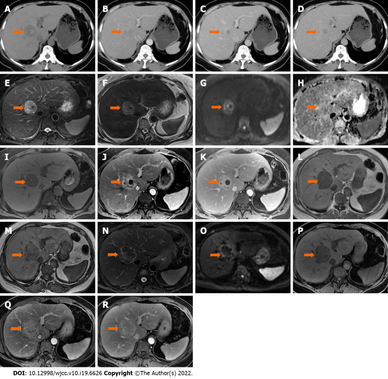
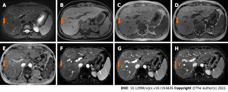
Three patients (one male and two females) were incidentally found to have a hepatic mass or nodule, without hepatomegaly or splenomegaly. Laboratory tests including liver function, serum hepatic tumor markers, and hepatitis serologic markers were normal. On MRI scans, all lesions showed lower signal intensity on in-phase images than on out-phase images. One case showed changes in signal intensity on T2 weighted images (WI) and diffusion WI, which shifted from hyperintensity to hypointensity with size enlargement between two rounds of imaging examination. These lesions exhibited different enhancement patterns on dynamic contrast enhancement series.
The MRI signal change and in-/out-phase image might provide useful information and help radiologists establish the diagnosis of intrahepatic extramedullary hematopoiesis.
肝内单独发生的髓外造血很少见,且易被误诊。关于该疾病的放射学文献均为病例报告。关于磁共振成像(MRI)作用的文献较少。最常用的成像方式是计算机断层扫描和超声。本报告旨在提供更多关于利用MRI观察髓外造血表现的数据,以帮助放射科医生进行诊断。
3例患者(1例男性和2例女性)偶然发现肝脏有肿块或结节,无肝肿大或脾肿大。包括肝功能、血清肝脏肿瘤标志物和肝炎血清学标志物在内的实验室检查均正常。在MRI扫描中,所有病变在同相位图像上的信号强度均低于反相位图像。1例在T2加权成像(WI)和扩散加权成像上显示信号强度变化,在两轮成像检查之间,随着大小增大,信号从高信号变为低信号。这些病变在动态对比增强序列上表现出不同的强化模式。
MRI信号变化及同/反相位图像可能提供有用信息,有助于放射科医生诊断肝内髓外造血。