• 文献检索
  • 文档翻译
  • 深度研究
  • 学术资讯
  • Suppr Zotero 插件Zotero 插件
  • 邀请有礼
  • 套餐&价格
  • 历史记录
应用&插件
Suppr Zotero 插件Zotero 插件浏览器插件Mac 客户端Windows 客户端微信小程序
定价
高级版会员购买积分包购买API积分包
服务
文献检索文档翻译深度研究API 文档MCP 服务
关于我们
关于 Suppr公司介绍联系我们用户协议隐私条款
关注我们

Suppr 超能文献

核心技术专利:CN118964589B侵权必究
粤ICP备2023148730 号-1Suppr @ 2026

文献检索

告别复杂PubMed语法,用中文像聊天一样搜索,搜遍4000万医学文献。AI智能推荐,让科研检索更轻松。

立即免费搜索

文件翻译

保留排版,准确专业,支持PDF/Word/PPT等文件格式,支持 12+语言互译。

免费翻译文档

深度研究

AI帮你快速写综述,25分钟生成高质量综述,智能提取关键信息,辅助科研写作。

立即免费体验

[F]氟胆碱正电子发射断层扫描/计算机断层扫描用于甲状旁腺成像的定量测量性能(P2TH研究)

Performance of quantitative measurements in [F]fluorocholine positron emission tomography/computed tomography for parathyroid imaging (P2TH study).

作者信息

Jacquet-Francillon Nicolas, Morelec Isabelle, Germain Natacha, Prades Jean-Michel, Habouzit Vincent, Mariat Christophe, Bonnefoy Pierre-Benoit, Prevot Nathalie

机构信息

Department of Nuclear Medicine, Saint-Étienne University Hospital, University of Saint-Étienne, Saint-Étienne, France.

Department of Nuclear Medicine, Hospices Civils de Lyon, Lyon, France.

出版信息

Front Med (Lausanne). 2022 Aug 2;9:956580. doi: 10.3389/fmed.2022.956580. eCollection 2022.

DOI:10.3389/fmed.2022.956580
PMID:35983092
原文链接:https://pmc.ncbi.nlm.nih.gov/articles/PMC9380568/
Abstract

OBJECTIVE

[F]Fluorocholine positron emission tomography/computed tomography (PET/CT) is used frequently in addition to [Tc]Tc-Sestamibi scintigraphy and ultrasonography for the location of hyperfunctioning parathyroid glands. The aim of this study is to evaluate the performance of quantitative criteria in [F]fluorocholine PET/CT for localization of hyperfunctioning parathyroid glands. The secondary objective is to highlight a correlation between the detection rate of [F]fluorocholine PET/CT and serum parathyroid hormone (PTH) level.

MATERIALS AND METHODS

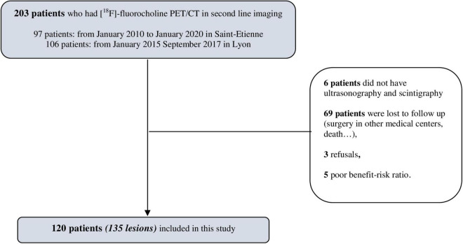
In two academic centers, we retrospectively included patients with biological hyperparathyroidism (HPT) and who had [F]fluorocholine PET/CT. After a visual analysis, to measure the overall performance of [F]fluorocholine PET/CT, a blind reading was carried out with standardized measurements of maximum standardized uptake value (SUV), liver ratio, thyroid ratio, and size ratio. We analyzed the quantitative criteria of [F]fluorocholine PET/CT compared to the histological results, in particular to identify differences between adenomas and hyperplasias. We compared the performance of each quantitative criterion to the overall sensitivity, specificity, positive predictive value (PPV), negative predictive value (NPV), and accuracy of [F]fluorocholine PET/CT. The detection rate of hyperfunctioning parathyroid glands was calculated in subgroups of serum PTH level.

RESULTS

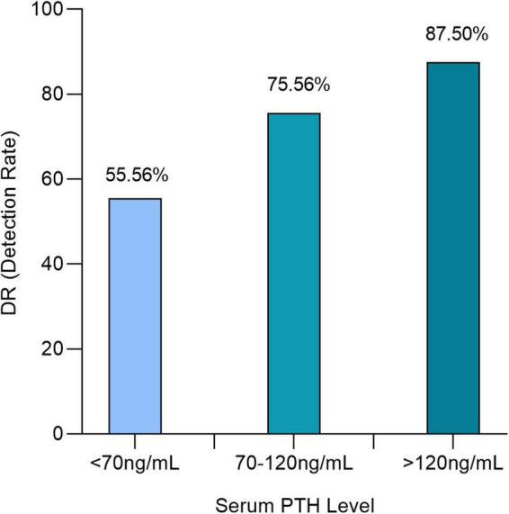
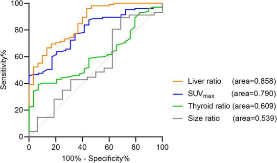
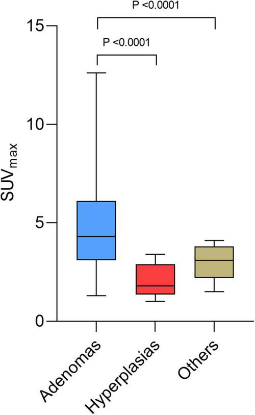
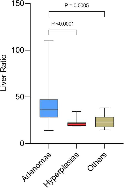
The quantitative criteria in [F]fluorocholine PET/CT were measured for 120 patients (135 lesions). The areas under the receiver operating characteristic (ROC) curve representing SUV and liver ratio were significantly increased. The optimal cut-off values represented by the maximum Youden index was >4.12 for SUV and >27.4 for liver ratio. Beyond certain threshold values of SUV (>4.12) or liver ratio (>38.1), all the lesions were histologically proven adenomas. SUV and liver ratio were significantly higher for adenomas than for hyperplasias and differential diagnosis ( = 0.0085 and = 0.0002). The positivity of [F]fluorocholine PET/CT was correlated with PTH level. Detection rates were 55.56, 75.56, and 87.5%, respectively, for serum PTH < 70, 70 to 120, and >120 ng/ml.

CONCLUSION

Semi-quantitative measurements (SUV and liver ratio) should be considered as additional tools in interpretation of [F]fluorocholine PET/CT. These quantitative parameters have lower overall performance but higher specificity than overall visual analysis in identifying an adenoma. Above certain threshold values, all lesions are adenomas. [F]fluorocholine PET/CT confirms excellent performance for the detection of hyperfunctional parathyroids. For serum PTH levels < 70 ng/ml, the detection rate of [F]fluorocholine PET/CT is strongly decreased.

摘要

目的

除了[锝]Tc - 甲氧基异丁基异腈闪烁扫描术和超声检查外,[氟]氟胆碱正电子发射断层扫描/计算机断层扫描(PET/CT)也经常用于定位功能亢进的甲状旁腺。本研究的目的是评估[氟]氟胆碱PET/CT定量标准在定位功能亢进甲状旁腺方面的性能。次要目的是突出[氟]氟胆碱PET/CT的检出率与血清甲状旁腺激素(PTH)水平之间的相关性。

材料与方法

在两个学术中心,我们回顾性纳入了患有生物性甲状旁腺功能亢进(HPT)且接受过[氟]氟胆碱PET/CT检查的患者。在视觉分析之后,为了测量[氟]氟胆碱PET/CT的整体性能,进行了盲法阅读,并对最大标准化摄取值(SUV)、肝脏比值、甲状腺比值和大小比值进行标准化测量。我们将[氟]氟胆碱PET/CT的定量标准与组织学结果进行了分析,特别是为了识别腺瘤和增生之间的差异。我们将每个定量标准的性能与[氟]氟胆碱PET/CT的整体敏感性、特异性、阳性预测值(PPV)、阴性预测值(NPV)和准确性进行了比较。在血清PTH水平的亚组中计算了功能亢进甲状旁腺的检出率。

结果

对120例患者(135个病灶)测量了[氟]氟胆碱PET/CT的定量标准。代表SUV和肝脏比值的受试者操作特征(ROC)曲线下面积显著增加。由最大约登指数表示的最佳截断值,SUV为>4.12,肝脏比值为>27.4。超过SUV(>4.12)或肝脏比值(>38.1)的某些阈值时,所有病灶经组织学证实为腺瘤。腺瘤的SUV和肝脏比值显著高于增生和鉴别诊断(P = 0.0085和P = 0.0002)。[氟]氟胆碱PET/CT的阳性与PTH水平相关。血清PTH<70、70至120和>120 ng/ml时,检出率分别为55.56%、75.56%和87.5%。

结论

在解读[氟]氟胆碱PET/CT时,应将半定量测量(SUV和肝脏比值)视为辅助工具。这些定量参数在识别腺瘤方面整体性能较低,但特异性高于整体视觉分析。超过某些阈值时,所有病灶均为腺瘤。[氟]氟胆碱PET/CT在检测功能亢进的甲状旁腺方面证实具有优异的性能。对于血清PTH水平<70 ng/ml,[氟]氟胆碱PET/CT的检出率会大幅降低。

https://cdn.ncbi.nlm.nih.gov/pmc/blobs/85e2/9380568/933e67e07e23/fmed-09-956580-g005.jpg
https://cdn.ncbi.nlm.nih.gov/pmc/blobs/85e2/9380568/80aa40bbf224/fmed-09-956580-g001.jpg
https://cdn.ncbi.nlm.nih.gov/pmc/blobs/85e2/9380568/9f81d240a540/fmed-09-956580-g002.jpg
https://cdn.ncbi.nlm.nih.gov/pmc/blobs/85e2/9380568/2270ca145a7d/fmed-09-956580-g003.jpg
https://cdn.ncbi.nlm.nih.gov/pmc/blobs/85e2/9380568/9768c7609287/fmed-09-956580-g004.jpg
https://cdn.ncbi.nlm.nih.gov/pmc/blobs/85e2/9380568/933e67e07e23/fmed-09-956580-g005.jpg

相似文献

1
Performance of quantitative measurements in [F]fluorocholine positron emission tomography/computed tomography for parathyroid imaging (P2TH study).[F]氟胆碱正电子发射断层扫描/计算机断层扫描用于甲状旁腺成像的定量测量性能(P2TH研究)
Front Med (Lausanne). 2022 Aug 2;9:956580. doi: 10.3389/fmed.2022.956580. eCollection 2022.
2
F-Fluorocholine PET/CT in the assessment of primary hyperparathyroidism compared with Tc-MIBI or Tc-tetrofosmin SPECT/CT: a prospective dual-centre study in 100 patients.氟代胆碱 PET/CT 与 Tc-MIBI 或 Tc-四氮茂 SPECT/CT 评估原发性甲状旁腺功能亢进症的比较:100 例患者的前瞻性双中心研究。
Eur J Nucl Med Mol Imaging. 2018 Sep;45(10):1762-1771. doi: 10.1007/s00259-018-3980-9. Epub 2018 Mar 8.
3
Diagnostic Performance of Cervical Ultrasound, Tc-Sestamibi Scintigraphy, and Contrast-Enhanced F-Fluorocholine PET in Primary Hyperparathyroidism.原发性甲状旁腺功能亢进症中颈超声、Tc-Sestamibi 闪烁扫描及 F-Fluorocholine PET 对比增强的诊断性能。
J Nucl Med. 2022 Jul;63(7):1081-1086. doi: 10.2967/jnumed.121.261900. Epub 2021 Dec 2.
4
Preoperative F-18 fluorocholine PET/CT for the detection of hyperfunctioning parathyroid glands in patients with secondary or tertiary hyperparathyroidism: comparison with Tc-99m sestamibi scan and neck ultrasound.术前 F-18 氟胆碱 PET/CT 检测继发性或三发性甲状旁腺功能亢进症患者的高功能甲状旁腺:与 Tc-99m sestamibi 扫描和颈部超声的比较。
Ann Nucl Med. 2020 Aug;34(8):527-537. doi: 10.1007/s12149-020-01479-2. Epub 2020 May 20.
5
Performance of F-18 Fluorocholine PET/CT for Detection of Hyperfunctioning Parathyroid Tissue in Patients with Elevated Parathyroid Hormone Levels and Negative or Discrepant Results in conventional Imaging.氟代胆碱 F-18 PET/CT 对甲状旁腺激素水平升高且常规影像学检查阴性或结果不一致的患者异位甲状旁腺组织的探测效能。
Korean J Radiol. 2020 Feb;21(2):236-247. doi: 10.3348/kjr.2019.0268.
6
A Pilot Comparison of 18F-fluorocholine PET/CT, Ultrasonography and 123I/99mTc-sestaMIBI Dual-Phase Dual-Isotope Scintigraphy in the Preoperative Localization of Hyperfunctioning Parathyroid Glands in Primary or Secondary Hyperparathyroidism: Influence of Thyroid Anomalies.18F-氟胆碱PET/CT、超声检查及123I/99mTc-甲氧基异丁基异腈双相双同位素闪烁扫描术在原发性或继发性甲状旁腺功能亢进症中功能亢进甲状旁腺术前定位的初步比较:甲状腺异常的影响
Medicine (Baltimore). 2015 Oct;94(41):e1701. doi: 10.1097/MD.0000000000001701.
7
Challenging pre-surgical localization of hyperfunctioning parathyroid glands in primary hyperparathyroidism: the added value of F-Fluorocholine PET/CT.原发性甲状旁腺功能亢进症中甲状旁腺功能亢进灶术前定位的挑战:F-氟代胆碱 PET/CT 的附加价值。
Eur J Nucl Med Mol Imaging. 2018 Sep;45(10):1772-1780. doi: 10.1007/s00259-018-4018-z. Epub 2018 Apr 22.
8
F-Fluorocholine-PET combined with contrast-enhanced CT for localizing hyperfunctioning parathyroid glands and optimizing surgical treatment in patients with hyperparathyroidism.氟代胆碱 PET 联合增强 CT 定位甲状旁腺功能亢进症并优化手术治疗。
Endocrine. 2022 Feb;75(2):593-600. doi: 10.1007/s12020-021-02877-w. Epub 2021 Sep 24.
9
Superior performance of 18F-fluorocholine digital PET/CT in the detection of parathyroid adenomas.18F-氟胆碱数字PET/CT在甲状旁腺腺瘤检测中的卓越性能。
Eur J Nucl Med Mol Imaging. 2020 Mar;47(3):572-578. doi: 10.1007/s00259-020-04680-7. Epub 2020 Jan 9.
10
F-Fluorocholine PET/CT Is More Sensitive Than C-Methionine PET/CT for the Localization of Hyperfunctioning Parathyroid Tissue in Primary Hyperparathyroidism.在原发性甲状旁腺功能亢进症中,F-氟胆碱PET/CT在定位功能亢进的甲状旁腺组织方面比C-蛋氨酸PET/CT更敏感。
J Nucl Med. 2022 May;63(5):785-791. doi: 10.2967/jnumed.121.262395. Epub 2021 Aug 19.

引用本文的文献

1
Digital versus analogue PET in parathyroid imaging: comparison of PET metrics and machine learning-based characterisation of hyperfunctioning lesions (the DIGI-PET study).甲状旁腺成像中的数字PET与模拟PET:PET指标比较及基于机器学习的功能亢进性病变特征分析(DIGI-PET研究)
Eur J Nucl Med Mol Imaging. 2025 Aug 22. doi: 10.1007/s00259-025-07508-4.

本文引用的文献

1
Histopathological Features of Parathyroid Adenoma and 18F-Choline Uptake in PET/MR of Primary Hyperparathyroidism.原发性甲状旁腺功能亢进症中甲状旁腺腺瘤的组织病理学特征及 18F-胆碱摄取的 PET/MR 表现
Clin Nucl Med. 2022 Feb 1;47(2):101-107. doi: 10.1097/RLU.0000000000003987.
2
The EANM practice guidelines for parathyroid imaging.欧洲核医学学会甲状旁腺影像实践指南。
Eur J Nucl Med Mol Imaging. 2021 Aug;48(9):2801-2822. doi: 10.1007/s00259-021-05334-y. Epub 2021 Apr 10.
3
Incidental Detection of a Small Cell Lung Cancer by 18F-Choline PET/CT Performed for Recurrent Hyperparathyroidism After Parathyroidectomy.
18F-胆碱 PET/CT 探测甲状旁腺癌术后复发性甲状旁腺功能亢进症时偶然发现小细胞肺癌
Clin Nucl Med. 2021 Feb 1;46(2):e109-e111. doi: 10.1097/RLU.0000000000003246.
4
Performance of F-18 Fluorocholine PET/CT for Detection of Hyperfunctioning Parathyroid Tissue in Patients with Elevated Parathyroid Hormone Levels and Negative or Discrepant Results in conventional Imaging.氟代胆碱 F-18 PET/CT 对甲状旁腺激素水平升高且常规影像学检查阴性或结果不一致的患者异位甲状旁腺组织的探测效能。
Korean J Radiol. 2020 Feb;21(2):236-247. doi: 10.3348/kjr.2019.0268.
5
Superior performance of 18F-fluorocholine digital PET/CT in the detection of parathyroid adenomas.18F-氟胆碱数字PET/CT在甲状旁腺腺瘤检测中的卓越性能。
Eur J Nucl Med Mol Imaging. 2020 Mar;47(3):572-578. doi: 10.1007/s00259-020-04680-7. Epub 2020 Jan 9.
6
F-Fluorocholine PET/CT in Primary Hyperparathyroidism: Superior Diagnostic Performance to Conventional Scintigraphic Imaging for Localization of Hyperfunctioning Parathyroid Glands.氟代胆碱正电子发射断层扫描/计算机断层扫描在原发性甲状旁腺功能亢进症中的应用:对异位甲状旁腺功能亢进的定位诊断优于传统显像。
J Nucl Med. 2020 Apr;61(4):577-583. doi: 10.2967/jnumed.119.229914. Epub 2019 Sep 27.
7
Parathyroid imaging with F-fluorocholine PET/CT as a first-line imaging modality in primary hyperparathyroidism: a retrospective cohort study.以F-氟胆碱PET/CT作为原发性甲状旁腺功能亢进一线成像方式的甲状旁腺成像:一项回顾性队列研究。
EJNMMI Res. 2019 Jul 31;9(1):72. doi: 10.1186/s13550-019-0544-3.
8
Choline PET/CT in parathyroid imaging: a systematic review.胆碱PET/CT在甲状旁腺成像中的应用:一项系统综述
Nucl Med Commun. 2019 Feb;40(2):96-105. doi: 10.1097/MNM.0000000000000952.
9
[Imaging of parathyroid adenomas with F‑18 choline PET-CT].[F-18胆碱PET-CT对甲状旁腺腺瘤的成像]
Wien Med Wochenschr. 2019 Feb;169(1-2):15-24. doi: 10.1007/s10354-018-0660-0. Epub 2018 Sep 27.
10
Additional value of integrated F-choline PET/4D contrast-enhanced CT in the localization of hyperfunctioning parathyroid glands and correlation with molecular profile.整合 F-胆碱 PET/4D 对比增强 CT 在高功能甲状旁腺定位中的附加价值及其与分子谱的相关性。
Eur J Nucl Med Mol Imaging. 2019 Mar;46(3):766-775. doi: 10.1007/s00259-018-4147-4. Epub 2018 Sep 15.