• 文献检索
  • 文档翻译
  • 深度研究
  • 学术资讯
  • Suppr Zotero 插件Zotero 插件
  • 邀请有礼
  • 套餐&价格
  • 历史记录
应用&插件
Suppr Zotero 插件Zotero 插件浏览器插件Mac 客户端Windows 客户端微信小程序
定价
高级版会员购买积分包购买API积分包
服务
文献检索文档翻译深度研究API 文档MCP 服务
关于我们
关于 Suppr公司介绍联系我们用户协议隐私条款
关注我们

Suppr 超能文献

核心技术专利:CN118964589B侵权必究
粤ICP备2023148730 号-1Suppr @ 2026

文献检索

告别复杂PubMed语法,用中文像聊天一样搜索,搜遍4000万医学文献。AI智能推荐,让科研检索更轻松。

立即免费搜索

文件翻译

保留排版,准确专业,支持PDF/Word/PPT等文件格式,支持 12+语言互译。

免费翻译文档

深度研究

AI帮你快速写综述,25分钟生成高质量综述,智能提取关键信息,辅助科研写作。

立即免费体验

对非视觉 arrestin 的研究揭示了功能位点之间的变构相互作用。

Surveying nonvisual arrestins reveals allosteric interactions between functional sites.

机构信息

Department of Biomedical Engineering, Case Western Reserve University, Cleveland, Ohio, USA.

Department of Biochemistry and Biophysics, University of Rochester, Rochester, New York, USA.

出版信息

Proteins. 2023 Jan;91(1):99-107. doi: 10.1002/prot.26413. Epub 2022 Aug 27.

DOI:10.1002/prot.26413
PMID:35988049
原文链接:https://pmc.ncbi.nlm.nih.gov/articles/PMC9771995/
Abstract

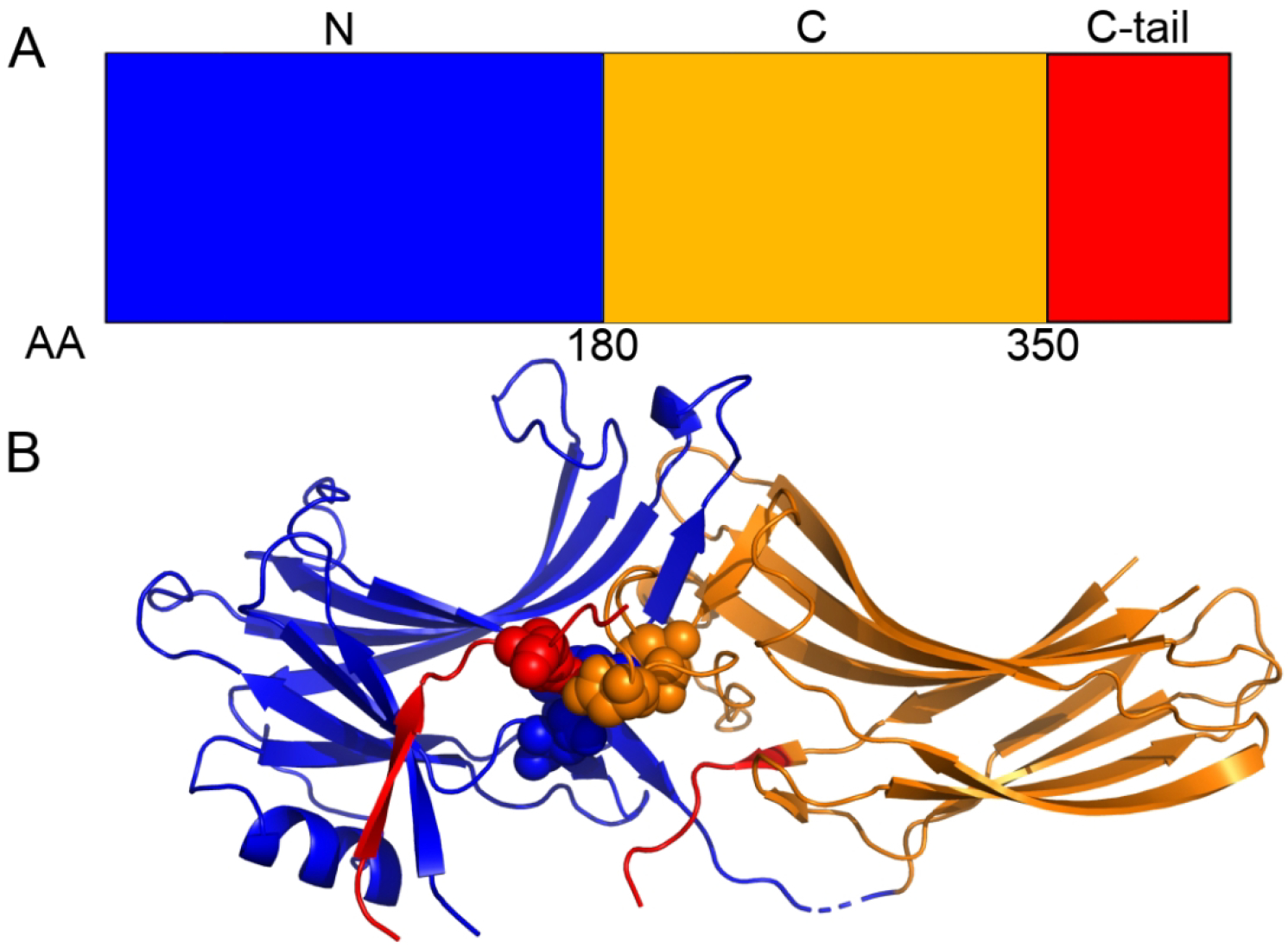
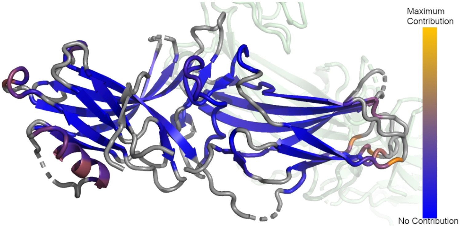
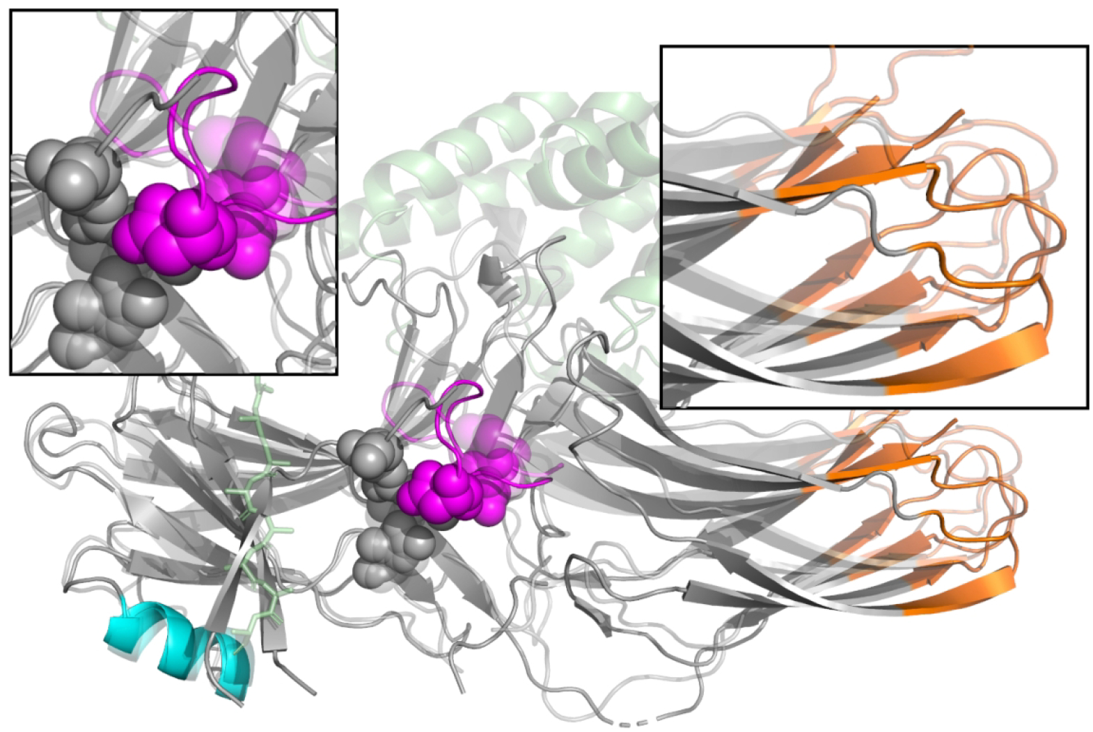
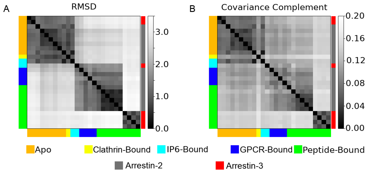
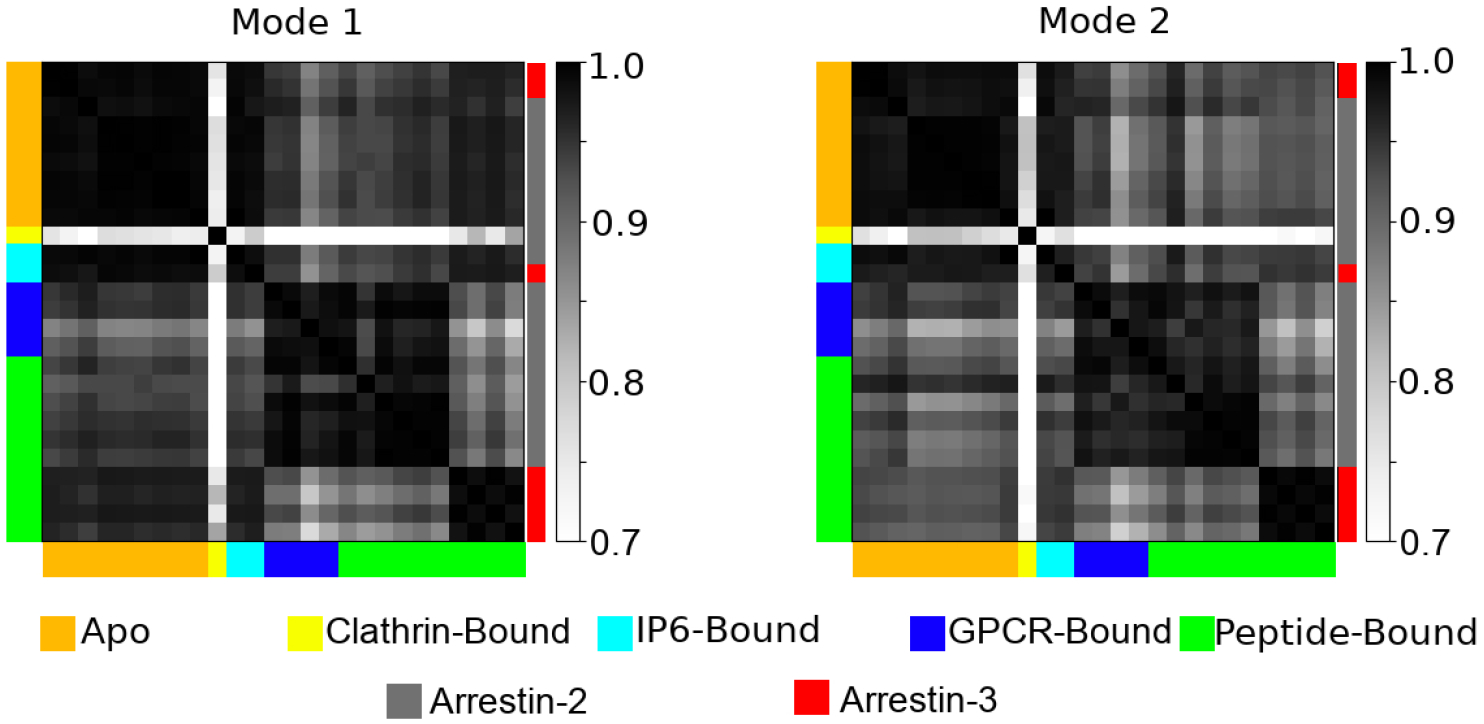
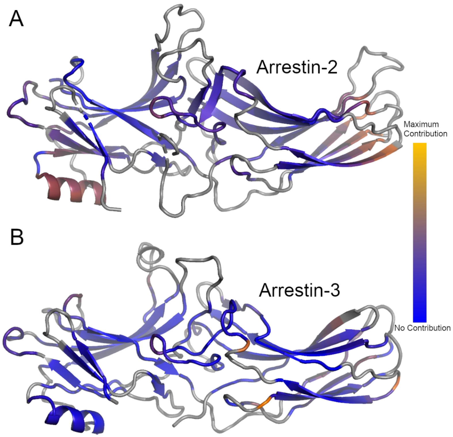
Arrestins are important scaffolding proteins that are expressed in all vertebrate animals. They regulate cell-signaling events upon binding to active G-protein coupled receptors (GPCR) and trigger endocytosis of active GPCRs. While many of the functional sites on arrestins have been characterized, the question of how these sites interact is unanswered. We used anisotropic network modeling (ANM) together with our covariance compliment techniques to survey all the available structures of the nonvisual arrestins to map how structural changes and protein-binding affect their structural dynamics. We found that activation and clathrin binding have a marked effect on arrestin dynamics, and that these dynamics changes are localized to a small number of distant functional sites. These sites include α-helix 1, the lariat loop, nuclear localization domain, and the C-domain β-sheets on the C-loop side. Our techniques suggest that clathrin binding and/or GPCR activation of arrestin perturb the dynamics of these sites independent of structural changes.

摘要

arrestins 是在所有脊椎动物中表达的重要支架蛋白。它们在与活性 G 蛋白偶联受体 (GPCR) 结合后调节细胞信号事件,并触发活性 GPCR 的内吞作用。虽然 arrestins 的许多功能位点已经得到了描述,但这些位点如何相互作用的问题仍未得到解答。我们使用各向异性网络建模 (ANM) 结合协方差互补技术,对所有可用的非视觉 arrestins 结构进行了调查,以绘制结构变化和蛋白质结合如何影响它们的结构动力学。我们发现,激活和网格蛋白结合对 arrestin 动力学有显著影响,并且这些动力学变化局限于少数几个遥远的功能位点。这些位点包括 α 螺旋 1、套索环、核定位域和 C 环侧的 C 结构域 β 片层。我们的技术表明,网格蛋白结合和/或 GPCR 激活 arrestin 会干扰这些位点的动力学,而与结构变化无关。

https://cdn.ncbi.nlm.nih.gov/pmc/blobs/c5ef/9771995/9e06728d5e38/nihms-1831167-f0006.jpg
https://cdn.ncbi.nlm.nih.gov/pmc/blobs/c5ef/9771995/432f7c0e8155/nihms-1831167-f0001.jpg
https://cdn.ncbi.nlm.nih.gov/pmc/blobs/c5ef/9771995/3ce708ccd581/nihms-1831167-f0002.jpg
https://cdn.ncbi.nlm.nih.gov/pmc/blobs/c5ef/9771995/ae0b5afed56a/nihms-1831167-f0003.jpg
https://cdn.ncbi.nlm.nih.gov/pmc/blobs/c5ef/9771995/4fd1e1f41c4e/nihms-1831167-f0004.jpg
https://cdn.ncbi.nlm.nih.gov/pmc/blobs/c5ef/9771995/d62cd7fc84f2/nihms-1831167-f0005.jpg
https://cdn.ncbi.nlm.nih.gov/pmc/blobs/c5ef/9771995/9e06728d5e38/nihms-1831167-f0006.jpg

相似文献

1
Surveying nonvisual arrestins reveals allosteric interactions between functional sites.对非视觉 arrestin 的研究揭示了功能位点之间的变构相互作用。
Proteins. 2023 Jan;91(1):99-107. doi: 10.1002/prot.26413. Epub 2022 Aug 27.
2
β-arrestins and G protein-coupled receptor trafficking.β-抑制蛋白与G蛋白偶联受体的转运
Handb Exp Pharmacol. 2014;219:173-86. doi: 10.1007/978-3-642-41199-1_9.
3
β-Arrestins and G protein-coupled receptor trafficking.β-抑制蛋白与G蛋白偶联受体的转运
Methods Enzymol. 2013;521:91-108. doi: 10.1016/B978-0-12-391862-8.00005-3.
4
Unraveling G protein-coupled receptor endocytosis pathways using real-time monitoring of agonist-promoted interaction between beta-arrestins and AP-2.利用实时监测激动剂促进的β-抑制蛋白与衔接蛋白AP-2之间的相互作用来揭示G蛋白偶联受体内吞途径
J Biol Chem. 2007 Oct 5;282(40):29089-100. doi: 10.1074/jbc.M700577200. Epub 2007 Aug 3.
5
β-Arrestin drives MAP kinase signalling from clathrin-coated structures after GPCR dissociation.G蛋白偶联受体解离后,β-抑制蛋白驱动网格蛋白包被结构的丝裂原活化蛋白激酶信号传导。
Nat Cell Biol. 2016 Mar;18(3):303-10. doi: 10.1038/ncb3307. Epub 2016 Feb 1.
6
Arrestin-Dependent and -Independent Internalization of G Protein-Coupled Receptors: Methods, Mechanisms, and Implications on Cell Signaling.G蛋白偶联受体的视 arrestin 依赖性和非依赖性内化:方法、机制及其对细胞信号传导的影响
Mol Pharmacol. 2021 Apr;99(4):242-255. doi: 10.1124/molpharm.120.000192. Epub 2021 Jan 20.
7
Discrete GPCR-triggered endocytic modes enable β-arrestins to flexibly regulate cell signaling.离散的 G 蛋白偶联受体触发的内吞模式使β-arrestin 能够灵活地调节细胞信号转导。
Elife. 2022 Oct 17;11:e81563. doi: 10.7554/eLife.81563.
8
beta-Arrestin/AP-2 interaction in G protein-coupled receptor internalization: identification of a beta-arrestin binging site in beta 2-adaptin.β-抑制蛋白/衔接蛋白-2相互作用在G蛋白偶联受体内化中的作用:β2-衔接蛋白中β-抑制蛋白结合位点的鉴定
J Biol Chem. 2002 Mar 15;277(11):9247-54. doi: 10.1074/jbc.M108490200. Epub 2002 Jan 2.
9
Membrane phosphoinositides regulate GPCR-β-arrestin complex assembly and dynamics.膜磷酯酰肌醇调节 G 蛋白偶联受体-β-arrestin 复合物的组装和动力学。
Cell. 2022 Nov 23;185(24):4560-4573.e19. doi: 10.1016/j.cell.2022.10.018. Epub 2022 Nov 10.
10
A beta-arrestin binding determinant common to the second intracellular loops of rhodopsin family G protein-coupled receptors.视紫红质家族G蛋白偶联受体第二细胞内环共有的β-抑制蛋白结合决定簇。
J Biol Chem. 2006 Feb 3;281(5):2932-8. doi: 10.1074/jbc.M508074200. Epub 2005 Nov 30.

引用本文的文献

1
Membrane phosphoinositides allosterically tune β-arrestin dynamics to facilitate GPCR core engagement.膜磷酸肌醇变构调节β-抑制蛋白的动力学,以促进与G蛋白偶联受体核心的结合。
bioRxiv. 2025 Jun 8:2025.06.06.658200. doi: 10.1101/2025.06.06.658200.
2
Association of Neurokinin-1 Receptor Signaling Pathways with Cancer.神经激肽-1 受体信号通路与癌症的关系。
Curr Med Chem. 2024;31(39):6460-6486. doi: 10.2174/0929867331666230818110812.

本文引用的文献

1
Structural studies of phosphorylation-dependent interactions between the V2R receptor and arrestin-2.V2R 受体和 arrestin-2 之间磷酸化依赖性相互作用的结构研究。
Nat Commun. 2021 Apr 22;12(1):2396. doi: 10.1038/s41467-021-22731-x.
2
Receptor-Arrestin Interactions: The GPCR Perspective.受体-抑制蛋白相互作用:G 蛋白偶联受体视角。
Biomolecules. 2021 Feb 4;11(2):218. doi: 10.3390/biom11020218.
3
RCSB Protein Data Bank: powerful new tools for exploring 3D structures of biological macromolecules for basic and applied research and education in fundamental biology, biomedicine, biotechnology, bioengineering and energy sciences.
RCSB 蛋白质数据库:用于基础生物学、生物医学、生物技术、生物工程和能源科学等领域的基础研究、应用研究和教育中探索生物大分子三维结构的强大新工具。
Nucleic Acids Res. 2021 Jan 8;49(D1):D437-D451. doi: 10.1093/nar/gkaa1038.
4
Lysine in the lariat loop of arrestins does not serve as phosphate sensor.衔接蛋白环赖氨酸不作为磷酸传感器。
J Neurochem. 2021 Feb;156(4):435-444. doi: 10.1111/jnc.15110. Epub 2020 Jul 11.
5
Molecular basis of β-arrestin coupling to formoterol-bound β-adrenoceptor.β-抑制蛋白与福莫特罗结合的β-肾上腺素能受体偶联的分子基础。
Nature. 2020 Jul;583(7818):862-866. doi: 10.1038/s41586-020-2419-1. Epub 2020 Jun 17.
6
Conformational Dynamics and Functional Implications of Phosphorylated β-Arrestins.磷酸化β-arrestin 的构象动态与功能意义
Structure. 2020 Mar 3;28(3):314-323.e3. doi: 10.1016/j.str.2019.12.008. Epub 2020 Jan 13.
7
Structure of the M2 muscarinic receptor-β-arrestin complex in a lipid nanodisc.M2 毒蕈碱型乙酰胆碱受体-β-arrestin 复合物在脂质纳米盘中的结构。
Nature. 2020 Mar;579(7798):297-302. doi: 10.1038/s41586-020-1954-0. Epub 2020 Jan 16.
8
Structure of the neurotensin receptor 1 in complex with β-arrestin 1.神经降压素受体 1 与β-arrestin 1 复合物的结构。
Nature. 2020 Mar;579(7798):303-308. doi: 10.1038/s41586-020-1953-1. Epub 2020 Jan 16.
9
Best Practices for Foundations in Molecular Simulations [Article v1.0].分子模拟基础的最佳实践 [文章v1.0]
Living J Comput Mol Sci. 2019;1(1). doi: 10.33011/livecoms.1.1.5957. Epub 2018 Nov 29.
10
A complex structure of arrestin-2 bound to a G protein-coupled receptor.一种与G蛋白偶联受体结合的视紫红质抑制蛋白-2的复杂结构。
Cell Res. 2019 Dec;29(12):971-983. doi: 10.1038/s41422-019-0256-2. Epub 2019 Nov 27.