Center for Neuroimmunology and Neuroinfectious Diseases, Washington University School of Medicine, St. Louis, MO, USA.
Department of Medicine, Washington University School of Medicine, St. Louis, MO, USA.
Brain. 2022 Dec 19;145(12):4193-4201. doi: 10.1093/brain/awac270.
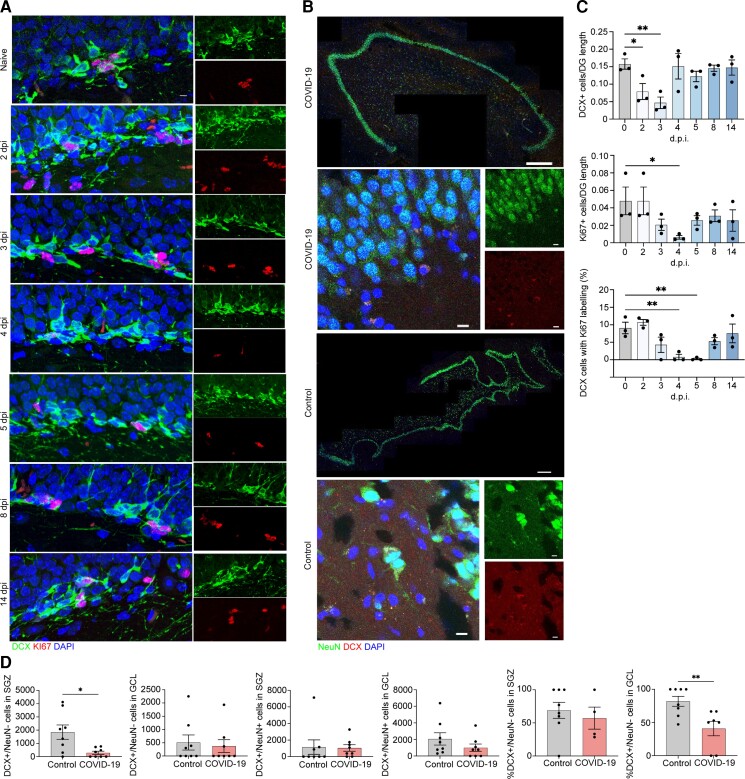
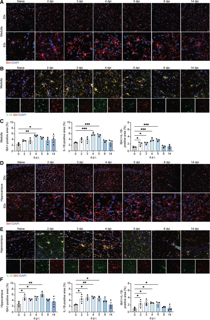
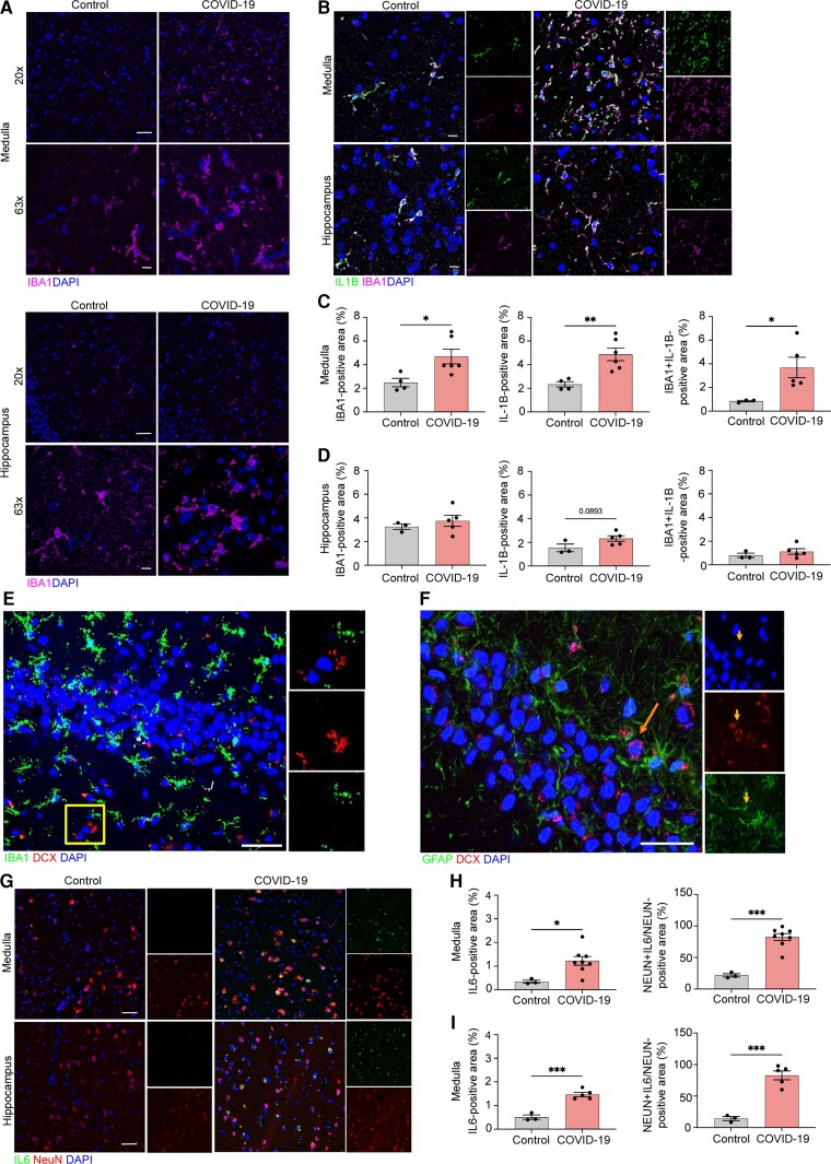
Infection with the severe acute respiratory syndrome coronavirus 2 (SARS-CoV-2) is associated with acute and postacute cognitive and neuropsychiatric symptoms including impaired memory, concentration, attention, sleep and affect. Mechanisms underlying these brain symptoms remain understudied. Here we report that SARS-CoV-2-infected hamsters exhibit a lack of viral neuroinvasion despite aberrant blood-brain barrier permeability. Hamsters and patients deceased from coronavirus disease 2019 (COVID-19) also exhibit microglial activation and expression of interleukin (IL)-1β and IL-6, especially within the hippocampus and the medulla oblongata, when compared with non-COVID control hamsters and humans who died from other infections, cardiovascular disease, uraemia or trauma. In the hippocampal dentate gyrus of both COVID-19 hamsters and humans, we observed fewer neuroblasts and immature neurons. Protracted inflammation, blood-brain barrier disruption and microglia activation may result in altered neurotransmission, neurogenesis and neuronal damage, explaining neuropsychiatric presentations of COVID-19. The involvement of the hippocampus may explain learning, memory and executive dysfunctions in COVID-19 patients.
严重急性呼吸综合征冠状病毒 2 (SARS-CoV-2) 的感染与急性和亚急性认知和神经精神症状有关,包括记忆力、注意力、注意力、睡眠和情绪受损。这些脑部症状的发病机制仍未得到充分研究。在这里,我们报告说,尽管血脑屏障通透性异常,但感染 SARS-CoV-2 的仓鼠并未出现病毒神经入侵。与非 COVID 对照组仓鼠和因其他感染、心血管疾病、尿毒症或创伤而死亡的人类相比,死于 2019 年冠状病毒病 (COVID-19) 的仓鼠和人类也表现出小胶质细胞激活和白细胞介素 (IL)-1β 和 IL-6 的表达,尤其是在海马体和延髓中。在 COVID-19 仓鼠和人类的海马齿状回中,我们观察到较少的神经前体细胞和未成熟神经元。持续的炎症、血脑屏障破坏和小胶质细胞激活可能导致神经递质、神经发生和神经元损伤的改变,从而解释了 COVID-19 的神经精神表现。海马体的参与可能解释了 COVID-19 患者的学习、记忆和执行功能障碍。