• 文献检索
  • 文档翻译
  • 深度研究
  • 学术资讯
  • Suppr Zotero 插件Zotero 插件
  • 邀请有礼
  • 套餐&价格
  • 历史记录
应用&插件
Suppr Zotero 插件Zotero 插件浏览器插件Mac 客户端Windows 客户端微信小程序
定价
高级版会员购买积分包购买API积分包
服务
文献检索文档翻译深度研究API 文档MCP 服务
关于我们
关于 Suppr公司介绍联系我们用户协议隐私条款
关注我们

Suppr 超能文献

核心技术专利:CN118964589B侵权必究
粤ICP备2023148730 号-1Suppr @ 2026

文献检索

告别复杂PubMed语法,用中文像聊天一样搜索,搜遍4000万医学文献。AI智能推荐,让科研检索更轻松。

立即免费搜索

文件翻译

保留排版,准确专业,支持PDF/Word/PPT等文件格式,支持 12+语言互译。

免费翻译文档

深度研究

AI帮你快速写综述,25分钟生成高质量综述,智能提取关键信息,辅助科研写作。

立即免费体验

Tbx18 通过招募上皮-间充质转化程序来协调心肌细胞的细胞结构转分化为起搏细胞。

Tbx18 Orchestrates Cytostructural Transdifferentiation of Cardiomyocytes to Pacemaker Cells by Recruiting the Epithelial-Mesenchymal Transition Program.

机构信息

Division of Cardiology, Department of Medicine, The Johns Hopkins University School of Medicine, Baltimore, Maryland 21205, United States.

Department of Pediatrics, Emory University, Atlanta, Georgia 30322, United States.

出版信息

J Proteome Res. 2022 Oct 7;21(10):2277-2292. doi: 10.1021/acs.jproteome.2c00133. Epub 2022 Aug 25.

DOI:10.1021/acs.jproteome.2c00133
PMID:36006872
原文链接:https://pmc.ncbi.nlm.nih.gov/articles/PMC9552783/
Abstract

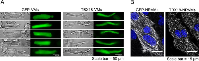
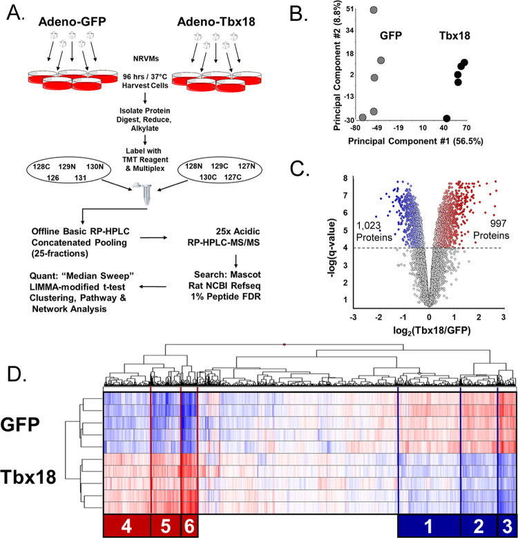
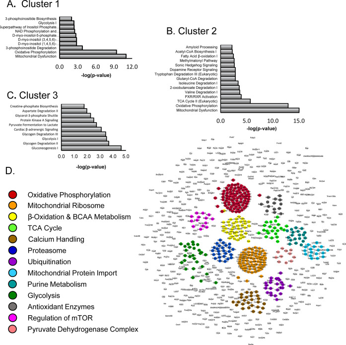
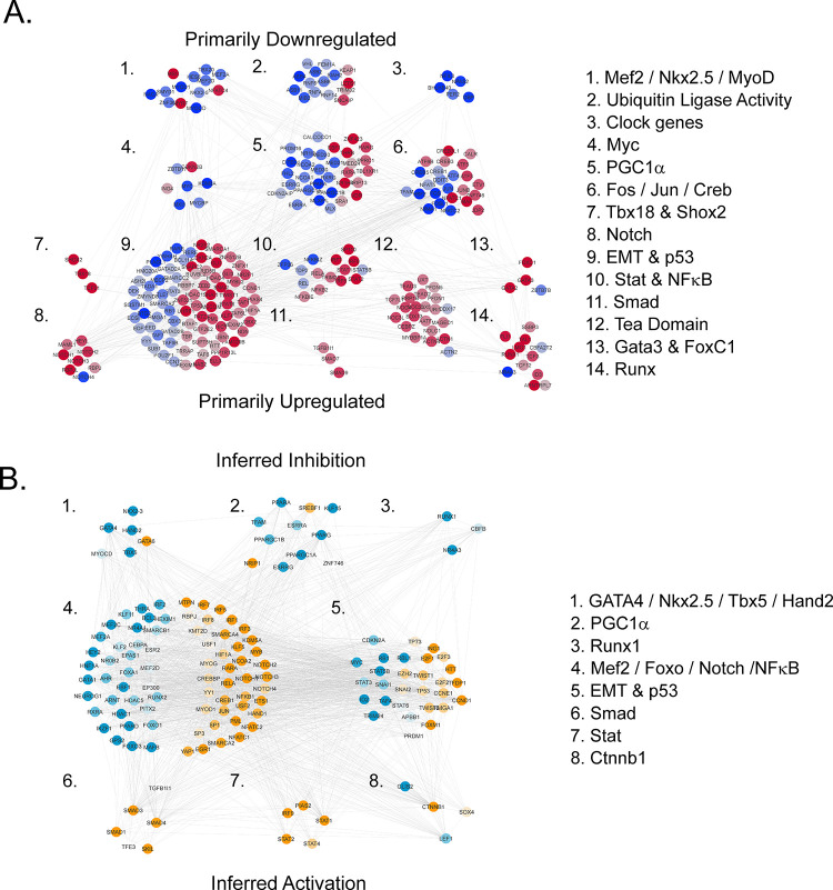
Previously, we reported that heterologous expression of an embryonic transcription factor, Tbx18, reprograms ventricular cardiomyocytes into induced pacemaker cells (Tbx18-iPMs), though the key pathways are unknown. Here, we have used a tandem mass tag proteomic approach to characterize the impact of Tbx18 on neonatal rat ventricular myocytes. Tbx18 expression triggered vast proteome remodeling. Tbx18-iPMs exhibited increased expression of known pacemaker ion channels, including Hcn4 and Cx45 as well as upregulation of the mechanosensitive ion channels Piezo1, Trpp2 (PKD2), and TrpM7. Metabolic pathways were broadly downregulated, as were ion channels associated with ventricular excitation-contraction coupling. Tbx18-iPMs also exhibited extensive intracellular cytoskeletal and extracellular matrix remodeling, including 96 differentially expressed proteins associated with the epithelial-to-mesenchymal transition (EMT). RNAseq extended coverage of low abundance transcription factors, revealing upregulation of EMT-inducing Snai1, Snai2, Twist1, Twist2, and Zeb2. Finally, network diffusion mapping of >200 transcriptional regulators indicates EMT and heart development factors occupy adjacent network neighborhoods downstream of Tbx18 but upstream of metabolic control factors. In conclusion, transdifferentiation of cardiac myocytes into pacemaker cells entails massive electrogenic, metabolic, and cytostructural remodeling. Structural changes exhibit hallmarks of the EMT. The results aid ongoing efforts to maximize the yield and phenotypic stability of engineered biological pacemakers.

摘要

先前,我们报道了胚胎转录因子 Tbx18 的异源表达可将心室肌细胞重编程为诱导性起搏细胞(Tbx18-iPMs),但其关键途径尚不清楚。在这里,我们使用串联质量标签蛋白质组学方法来研究 Tbx18 对新生大鼠心室肌细胞的影响。Tbx18 的表达引发了广泛的蛋白质组重编程。Tbx18-iPMs 表现出已知起搏离子通道的表达增加,包括 Hcn4 和 Cx45,以及机械敏感离子通道 Piezo1、Trpp2(PKD2)和 TrpM7 的上调。代谢途径广泛下调,与心室兴奋-收缩偶联相关的离子通道也是如此。Tbx18-iPMs 还表现出广泛的细胞内细胞骨架和细胞外基质重塑,包括 96 种与上皮-间质转化(EMT)相关的差异表达蛋白。RNAseq 扩展了低丰度转录因子的覆盖范围,揭示了 EMT 诱导因子 Snai1、Snai2、Twist1、Twist2 和 Zeb2 的上调。最后,>200 个转录调节剂的网络扩散映射表明,EMT 和心脏发育因子占据 Tbx18 下游的相邻网络邻域,但位于代谢控制因子的上游。总之,心肌细胞向起搏细胞的转分化需要大量的电生成、代谢和细胞结构重塑。结构变化表现出 EMT 的特征。这些结果有助于正在进行的努力,以最大限度地提高工程生物起搏器的产量和表型稳定性。

https://cdn.ncbi.nlm.nih.gov/pmc/blobs/f33c/9552783/f6ee92b574d5/pr2c00133_0008.jpg
https://cdn.ncbi.nlm.nih.gov/pmc/blobs/f33c/9552783/486889228f80/pr2c00133_0002.jpg
https://cdn.ncbi.nlm.nih.gov/pmc/blobs/f33c/9552783/c58f7e94ea8b/pr2c00133_0003.jpg
https://cdn.ncbi.nlm.nih.gov/pmc/blobs/f33c/9552783/6921d4f92ffc/pr2c00133_0004.jpg
https://cdn.ncbi.nlm.nih.gov/pmc/blobs/f33c/9552783/bf5c605cc528/pr2c00133_0005.jpg
https://cdn.ncbi.nlm.nih.gov/pmc/blobs/f33c/9552783/916c87b1644d/pr2c00133_0006.jpg
https://cdn.ncbi.nlm.nih.gov/pmc/blobs/f33c/9552783/3906ee57622f/pr2c00133_0007.jpg
https://cdn.ncbi.nlm.nih.gov/pmc/blobs/f33c/9552783/f6ee92b574d5/pr2c00133_0008.jpg

相似文献

1
Tbx18 Orchestrates Cytostructural Transdifferentiation of Cardiomyocytes to Pacemaker Cells by Recruiting the Epithelial-Mesenchymal Transition Program.Tbx18 通过招募上皮-间充质转化程序来协调心肌细胞的细胞结构转分化为起搏细胞。
J Proteome Res. 2022 Oct 7;21(10):2277-2292. doi: 10.1021/acs.jproteome.2c00133. Epub 2022 Aug 25.
2
Induced cardiac pacemaker cells survive metabolic stress owing to their low metabolic demand.诱导性心脏起搏器细胞因其低代谢需求而能在代谢应激中存活。
Exp Mol Med. 2019 Sep 13;51(9):1-12. doi: 10.1038/s12276-019-0303-6.
3
TBX18 overexpression enhances pacemaker function in a rat subsidiary atrial pacemaker model of sick sinus syndrome.TBX18 过表达增强病态窦房结综合征大鼠副房起博器模型中的起博功能。
J Physiol. 2018 Dec;596(24):6141-6155. doi: 10.1113/JP276508. Epub 2018 Oct 13.
4
TBX18 gene induces adipose-derived stem cells to differentiate into pacemaker-like cells in the myocardial microenvironment.TBX18基因在心肌微环境中诱导脂肪来源干细胞分化为起搏样细胞。
Int J Mol Med. 2016 Nov;38(5):1403-1410. doi: 10.3892/ijmm.2016.2736. Epub 2016 Sep 14.
5
Functional biological pacemaker generation by T-Box18 protein expression via stem cell and viral delivery approaches in a murine model of complete heart block.通过干细胞和病毒传递方法在完全性心脏传导阻滞的小鼠模型中表达 T-Box18 蛋白生成功能性生物起搏器。
Pharmacol Res. 2019 Mar;141:443-450. doi: 10.1016/j.phrs.2019.01.034. Epub 2019 Jan 21.
6
Transcription Factor Reprograms Vascular Smooth Muscle Cells of Ascending Aorta to Pacemaker-Like Cells.转录因子将升主动脉血管平滑肌细胞重编程为起搏样细胞。
DNA Cell Biol. 2019 Dec;38(12):1470-1479. doi: 10.1089/dna.2019.4940. Epub 2019 Oct 18.
7
Pacemaker cell characteristics of differentiated and HCN4-transduced human mesenchymal stem cells.分化的和转染 HCN4 的人间质干细胞的起搏器细胞特征。
Life Sci. 2019 Sep 1;232:116620. doi: 10.1016/j.lfs.2019.116620. Epub 2019 Jul 7.
8
Transcription factor TBX18 promotes adult rat bone mesenchymal stem cell differentiation to biological pacemaker cells.转录因子 TBX18 促进成年大鼠骨髓间充质干细胞向生物起搏器细胞的分化。
Int J Mol Med. 2018 Feb;41(2):845-851. doi: 10.3892/ijmm.2017.3259. Epub 2017 Nov 16.
9
Transcriptional suppression of connexin43 by TBX18 undermines cell-cell electrical coupling in postnatal cardiomyocytes.转录抑制因子 TBX18 抑制了出生后心肌细胞缝隙连接蛋白 43 的表达,破坏了细胞间的电耦联。
J Biol Chem. 2011 Apr 22;286(16):14073-9. doi: 10.1074/jbc.M110.185298. Epub 2011 Jan 4.
10
Tbx18 promoted the conversion of human-induced pluripotent stem cell-derived cardiomyocytes into sinoatrial node-like pacemaker cells.Tbx18 促进人诱导多能干细胞衍生的心肌细胞向窦房结样起搏细胞的转化。
Cell Biol Int. 2022 Mar;46(3):403-414. doi: 10.1002/cbin.11738. Epub 2021 Dec 30.

引用本文的文献

1
Inhibition of Cardiac p38 Highlights the Role of the Phosphoproteome in Heart Failure Progression.抑制心脏中的p38突出了磷酸化蛋白质组在心力衰竭进展中的作用。
ACS Omega. 2025 Aug 6;10(32):36082-36097. doi: 10.1021/acsomega.5c03687. eCollection 2025 Aug 19.
2
Inhibition of Cardiac p38 Highlights the Role of the Phosphoproteome in Heart Failure Progression.抑制心脏中的p38凸显了磷酸化蛋白质组在心力衰竭进展中的作用。
bioRxiv. 2024 Nov 20:2024.11.20.624554. doi: 10.1101/2024.11.20.624554.
3
Proteomics of the heart.心脏蛋白质组学。

本文引用的文献

1
Quantitative proteomics characterization of acutely isolated primary adult rat cardiomyocytes and fibroblasts.定量蛋白质组学分析急性分离的成年大鼠原代心肌细胞和成纤维细胞。
J Mol Cell Cardiol. 2020 Jun;143:63-70. doi: 10.1016/j.yjmcc.2020.04.021. Epub 2020 Apr 20.
2
Quantitative proteomics and single-nucleus transcriptomics of the sinus node elucidates the foundation of cardiac pacemaking.窦房结的定量蛋白质组学和单核转录组学阐明了心脏起搏的基础。
Nat Commun. 2019 Jun 28;10(1):2889. doi: 10.1038/s41467-019-10709-9.
3
Endothelial to Mesenchymal Transition in Cardiovascular Disease: JACC State-of-the-Art Review.
Physiol Rev. 2024 Jul 1;104(3):931-982. doi: 10.1152/physrev.00026.2023. Epub 2024 Feb 1.
4
Identification of RNA reads encoding different channels in isolated rat ventricular myocytes and the effect of cell stretching on L-type Cacurrent.鉴定分离的大鼠心室肌细胞中不同通道的 RNA 读取,以及细胞拉伸对 L 型钙电流的影响。
Biol Direct. 2023 Oct 30;18(1):70. doi: 10.1186/s13062-023-00427-0.
5
Harnessing cell reprogramming for cardiac biological pacing.利用细胞重编程实现心脏生物起搏。
J Biomed Sci. 2023 Aug 26;30(1):74. doi: 10.1186/s12929-023-00970-y.
心血管疾病中的内皮细胞向间充质细胞转化:JACC 前沿综述。
J Am Coll Cardiol. 2019 Jan 22;73(2):190-209. doi: 10.1016/j.jacc.2018.09.089.
4
STRING v11: protein-protein association networks with increased coverage, supporting functional discovery in genome-wide experimental datasets.STRING v11:具有增强覆盖范围的蛋白质-蛋白质相互作用网络,支持在全基因组实验数据集的功能发现。
Nucleic Acids Res. 2019 Jan 8;47(D1):D607-D613. doi: 10.1093/nar/gky1131.
5
Cytoscape StringApp: Network Analysis and Visualization of Proteomics Data.Cytoscape StringApp:蛋白质组学数据的网络分析和可视化。
J Proteome Res. 2019 Feb 1;18(2):623-632. doi: 10.1021/acs.jproteome.8b00702. Epub 2018 Dec 5.
6
The PRIDE database and related tools and resources in 2019: improving support for quantification data.PRIDE 数据库及相关工具和资源在 2019 年的进展:提高定量数据支持。
Nucleic Acids Res. 2019 Jan 8;47(D1):D442-D450. doi: 10.1093/nar/gky1106.
7
Proteomic analysis identifies transcriptional cofactors and homeobox transcription factors as TBX18 binding proteins.蛋白质组学分析鉴定出转录共因子和同源盒转录因子为 TBX18 结合蛋白。
PLoS One. 2018 Aug 2;13(8):e0200964. doi: 10.1371/journal.pone.0200964. eCollection 2018.
8
Polycystin-2-dependent control of cardiomyocyte autophagy.多囊蛋白-2 依赖性调控心肌细胞自噬。
J Mol Cell Cardiol. 2018 May;118:110-121. doi: 10.1016/j.yjmcc.2018.03.002. Epub 2018 Mar 6.
9
Piezo1 channels sense whole body physical activity to reset cardiovascular homeostasis and enhance performance.Piezo1通道感知全身身体活动,以重置心血管稳态并提高运动表现。
Nat Commun. 2017 Aug 24;8(1):350. doi: 10.1038/s41467-017-00429-3.
10
Stretch-activated channel Piezo1 is up-regulated in failure heart and cardiomyocyte stimulated by AngII.机械敏感性通道Piezo1在衰竭心脏和受血管紧张素II刺激的心肌细胞中上调。
Am J Transl Res. 2017 Jun 15;9(6):2945-2955. eCollection 2017.