Feher Balazs, Kuchler Ulrike, Schwendicke Falk, Schneider Lisa, Cejudo Grano de Oro Jose Eduardo, Xi Tong, Vinayahalingam Shankeeth, Hsu Tzu-Ming Harry, Brinz Janet, Chaurasia Akhilanand, Dhingra Kunaal, Gaudin Robert Andre, Mohammad-Rahimi Hossein, Pereira Nielsen, Perez-Pastor Francesc, Tryfonos Olga, Uribe Sergio E, Hanisch Marcel, Krois Joachim
Department of Oral Surgery, University Clinic of Dentistry, Medical University of Vienna, 1090 Vienna, Austria.
Competence Center Oral Biology, University Clinic of Dentistry, Medical University of Vienna, 1090 Vienna, Austria.
Diagnostics (Basel). 2022 Aug 14;12(8):1968. doi: 10.3390/diagnostics12081968.
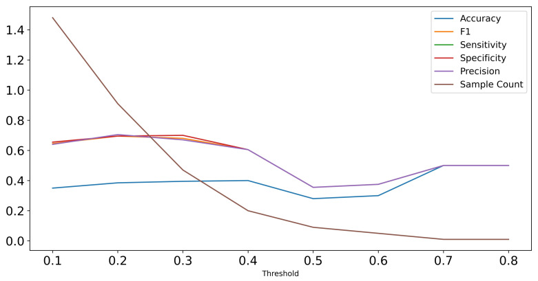
The detection and classification of cystic lesions of the jaw is of high clinical relevance and represents a topic of interest in medical artificial intelligence research. The human clinical diagnostic reasoning process uses contextual information, including the spatial relation of the detected lesion to other anatomical structures, to establish a preliminary classification. Here, we aimed to emulate clinical diagnostic reasoning step by step by using a combined object detection and image segmentation approach on panoramic radiographs (OPGs). We used a multicenter training dataset of 855 OPGs (all positives) and an evaluation set of 384 OPGs (240 negatives). We further compared our models to an international human control group of ten dental professionals from seven countries. The object detection model achieved an average precision of 0.42 (intersection over union (IoU): 0.50, maximal detections: 100) and an average recall of 0.394 (IoU: 0.50-0.95, maximal detections: 100). The classification model achieved a sensitivity of 0.84 for odontogenic cysts and 0.56 for non-odontogenic cysts as well as a specificity of 0.59 for odontogenic cysts and 0.84 for non-odontogenic cysts (IoU: 0.30). The human control group achieved a sensitivity of 0.70 for odontogenic cysts, 0.44 for non-odontogenic cysts, and 0.56 for OPGs without cysts as well as a specificity of 0.62 for odontogenic cysts, 0.95 for non-odontogenic cysts, and 0.76 for OPGs without cysts. Taken together, our results show that a combined object detection and image segmentation approach is feasible in emulating the human clinical diagnostic reasoning process in classifying cystic lesions of the jaw.
颌骨囊性病变的检测与分类具有高度的临床相关性,是医学人工智能研究中一个备受关注的课题。人类临床诊断推理过程利用上下文信息,包括检测到的病变与其他解剖结构的空间关系,来进行初步分类。在此,我们旨在通过对全景X线片(OPG)使用目标检测与图像分割相结合的方法,逐步模拟临床诊断推理过程。我们使用了一个包含855张OPG的多中心训练数据集(均为阳性)和一个包含384张OPG的评估集(240张为阴性)。我们还将我们的模型与来自七个国家的十名牙科专业人员组成的国际人类对照组进行了比较。目标检测模型的平均精度为0.42(交并比(IoU):0.50,最大检测数:100),平均召回率为0.394(IoU:0.50 - 0.95,最大检测数:100)。分类模型对牙源性囊肿的敏感度为0.84,对非牙源性囊肿的敏感度为0.56,对牙源性囊肿的特异度为0.59,对非牙源性囊肿的特异度为0.84(IoU:0.30)。人类对照组对牙源性囊肿的敏感度为0.70,对非牙源性囊肿的敏感度为0.44,对无囊肿的OPG的敏感度为0.56,对牙源性囊肿的特异度为0.62,对非牙源性囊肿的特异度为0.95,对无囊肿的OPG的特异度为0.76。综上所述,我们的结果表明,目标检测与图像分割相结合的方法在模拟人类临床诊断推理过程以分类颌骨囊性病变方面是可行的。