Department of Pathology, Microbiology, and Immunology, School of Veterinary Medicine, University of California, Davis, Davis, CA, USA.
Sci Rep. 2022 Aug 26;12(1):14578. doi: 10.1038/s41598-022-18771-y.
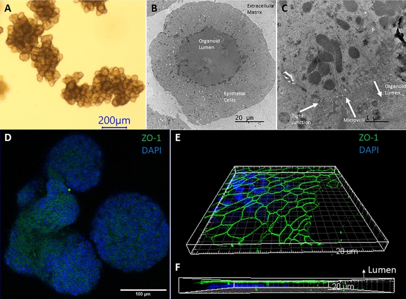
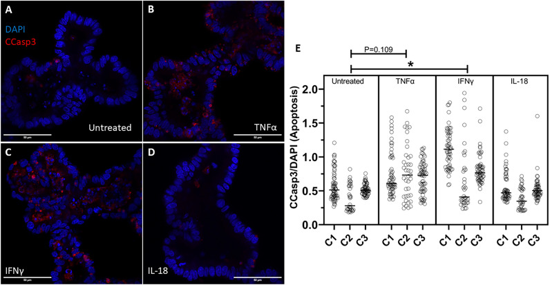
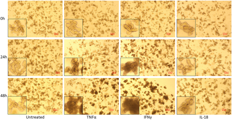
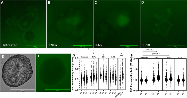
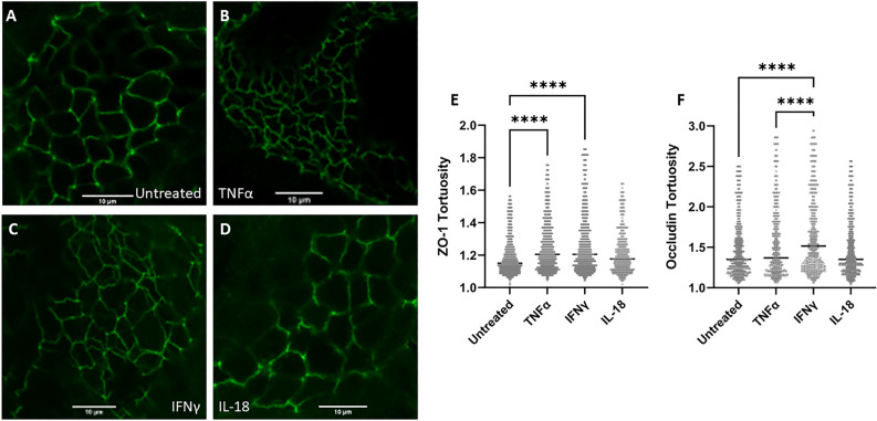
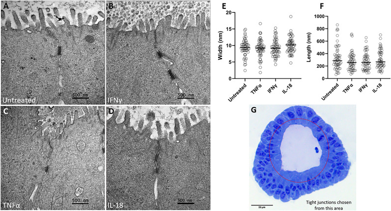
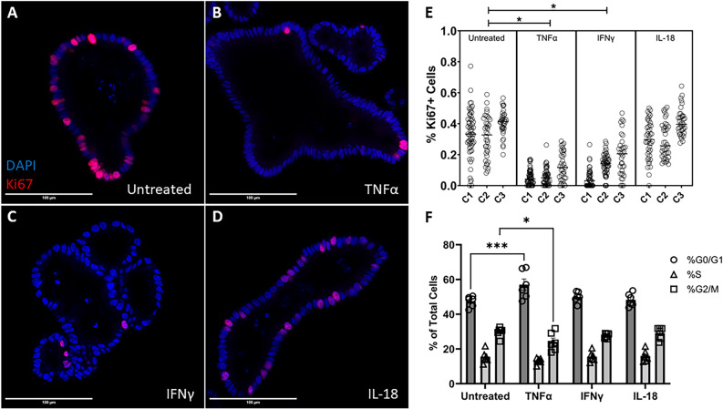
The small intestinal mucosa constitutes a physical barrier separating the gut lumen from sterile internal tissues. Junctional complexes between cells regulate transport across the barrier, preventing water loss and the entry of noxious molecules or pathogens. Inflammatory diseases in cattle disrupt this barrier; nonetheless, mechanisms of barrier disruption in cattle are poorly understood. We investigated the direct effects of three inflammatory cytokines, TNFα, IFNγ, and IL-18, on the bovine intestinal barrier utilizing intestinal organoids. Flux of fluorescein isothiocyanate (FITC)-labeled dextran was used to investigate barrier permeability. Immunocytochemistry and transmission electron microscopy were used to investigate junctional morphology, specifically tortuosity and length/width, respectively. Immunocytochemistry and flow cytometry was used to investigate cellular turnover via proliferation and apoptosis. Our study shows that 24-h cytokine treatment with TNFα or IFNγ significantly increased dextran permeability and tight junctional tortuosity, and reduced cellular proliferation. TNFα reduced the percentage of G2/M phase cells, and IFNγ treatment increased cell apoptotic rate. IL-18 did not directly induce significant changes to barrier permeability or cellular turnover. Our study concludes that the inflammatory cytokines, TNFα and IFNγ, directly induce intestinal epithelial barrier dysfunction and alter the tight junctional morphology and rate of cellular turnover in bovine intestinal epithelial cells.
小肠黏膜构成了一个物理屏障,将肠道腔与无菌的内部组织隔开。细胞之间的连接复合体调节着屏障的转运,防止水分流失和有害分子或病原体的进入。牛的炎症性疾病会破坏这种屏障;然而,牛的屏障破坏机制还知之甚少。我们利用肠类器官研究了三种炎症细胞因子 TNFα、IFNγ 和 IL-18 对牛肠道屏障的直接影响。使用荧光素异硫氰酸酯(FITC)标记的葡聚糖来研究屏障通透性。免疫细胞化学和透射电子显微镜分别用于研究连接形态,特别是扭曲度和长度/宽度。免疫细胞化学和流式细胞术用于通过增殖和细胞凋亡研究细胞更新。我们的研究表明,TNFα 或 IFNγ 的 24 小时细胞因子处理显著增加了葡聚糖的通透性和紧密连接的扭曲度,并减少了细胞增殖。TNFα 降低了 G2/M 期细胞的百分比,IFNγ 处理增加了细胞凋亡率。IL-18 并未直接诱导屏障通透性或细胞更新发生显著变化。我们的研究得出结论,炎症细胞因子 TNFα 和 IFNγ 直接诱导肠道上皮屏障功能障碍,并改变牛肠道上皮细胞中紧密连接的形态和细胞更新率。