Yoshida Shino, Nakazawa Meg, Kawasaki Minae, Ambrosini Yoko M
Department of Veterinary Clinical Sciences, College of Veterinary Medicine, Washington State University, Pullman, WA, United States.
Front Vet Sci. 2024 Dec 18;11:1483421. doi: 10.3389/fvets.2024.1483421. eCollection 2024.
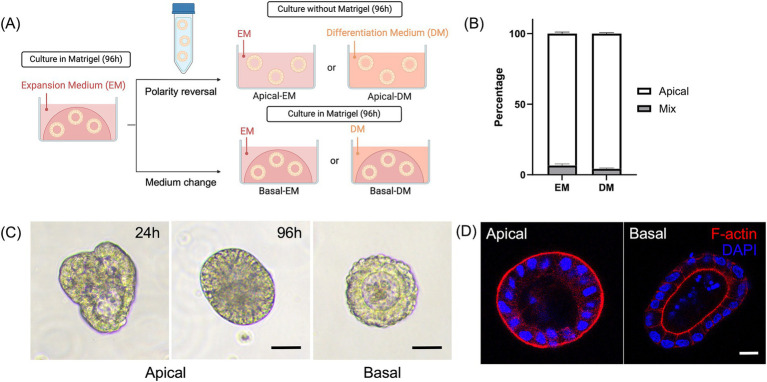
Dogs are increasingly recognized as valuable large animal models for understanding human intestinal diseases, as they naturally develop conditions similar to those in humans, such as Enterohemorrhagic , , inflammatory bowel disease, and ulcerative colitis. Given the similarity in gut flora between dogs and humans, canine intestinal models are ideal for translational research. However, conventional extracellular matrix-embedded organoids present challenges in accessing the lumen, which is critical for gut function. This study aimed to investigate the feasibility of inducing polarity reversal and differentiation in canine apical-out colonic organoids (colonoids), evaluate their barrier integrity, and visualize host-pathogen interactions. Our results demonstrated successful polarity reversal and differentiation induction while maintaining barrier integrity. Polarity reversal allowed for enhanced observation of host-pathogen interactions, facilitating visual assessments and membrane integrity evaluations using both pathogenic and nonpathogenic . This process led to the downregulation of stem cell marker and upregulation of intestinal epithelial cell marker , indicating differentiation. Further differentiation was observed with the use of a differentiation culture medium, resulting in significant upregulation of and goblet cell marker . The findings suggest that apical-out canine colonoids can serve as physiologic and valuable models for studying the pathogenic mechanisms and clinical significance of intestinal diseases in dogs. This model has the potential to advance both canine and human gastrointestinal research, enhancing our understanding of gastrointestinal physiology and pathology and aiding in the development of novel therapeutics.
狗越来越被认为是理解人类肠道疾病的有价值的大型动物模型,因为它们自然会发展出与人类相似的病症,如肠出血性疾病、炎症性肠病和溃疡性结肠炎。鉴于狗和人类肠道菌群的相似性,犬肠道模型是转化研究的理想选择。然而,传统的细胞外基质包埋类器官在进入管腔方面存在挑战,而管腔对肠道功能至关重要。本研究旨在探讨诱导犬顶出式结肠类器官(结肠小体)极性逆转和分化的可行性,评估其屏障完整性,并可视化宿主-病原体相互作用。我们的结果表明,在保持屏障完整性的同时成功诱导了极性逆转和分化。极性逆转有助于增强对宿主-病原体相互作用的观察,便于使用致病性和非致病性[具体内容缺失]进行视觉评估和膜完整性评估。这一过程导致干细胞标志物[具体内容缺失]下调,肠道上皮细胞标志物[具体内容缺失]上调,表明发生了分化。使用分化培养基观察到进一步的分化,导致[具体内容缺失]和杯状细胞标志物[具体内容缺失]显著上调。研究结果表明,顶出式犬结肠小体可作为研究犬肠道疾病致病机制和临床意义的生理学和有价值的模型。该模型有潜力推动犬类和人类胃肠道研究的发展,增进我们对胃肠道生理学和病理学的理解,并有助于开发新的治疗方法。