Sharawy Nivin, Imam Ahmad Abdel-Aliem, Aboulhoda Basma Emad, Khalifa Mohamed Mansour, Morcos George N B, Abd Algaleel Waleed Ahmed, Moustafa Passant E, Abdelbaset Marwan A, Shoukry Tarek
Department of Physiology, Faculty of Medicine, Cairo University, Cairo, Egypt.
Preclinical Sciences, College of Osteopathic Medicine, William Carey University, Hattiesburg, MS, United States.
Front Physiol. 2022 Aug 12;13:953206. doi: 10.3389/fphys.2022.953206. eCollection 2022.
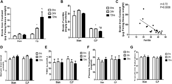
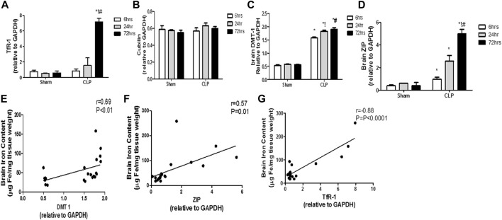
Encephalopathy is a frequent and lethal consequence of sepsis. Recently, a growing body of evidence has provided important insights into the role of iron dyshomeostasis in the context of inflammation. The molecular mechanisms underlying iron dyshomeostasis and its relationship with macrophage phenotypes are largely unknown. Here, we aimed to characterize the changes in iron-transporter and storage proteins and the microglia phenotype that occur during the course of sepsis, as well as their relationship with sepsis-induced encephalopathy. We used a cecal ligation and puncture (CLP) murine model that closely resembles sepsis-induced encephalopathy. Rats were subjected to CLP or sham laparotomy, then were neurologically assessed at 6 h, 24 h, and 3 days after sepsis induction. The serum and brain were collected for subsequent biochemical, histological, and immunohistochemical assessment. Here, an iron excess was observed at time points that followed the pro-inflammatory macrophage polarization in CLP-induced encephalopathy. Our results revealed that the upregulation of non-transferrin-bound iron uptake (NTBI) and ferritin reduction appeared to be partially responsible for the excess free iron detected within the brain tissues. We further demonstrated that the microglia were shifted toward the pro-inflammatory phenotype, leading to persistent neuro-inflammation and neuronal damage after CLP. Taken together, these findings led us to conclude that sepsis increased the susceptibility of the brain to the iron burden the upregulation of NTBI and the reduction of ferritin, which was concomitantly and correlatively associated with dominance of pro-inflammatory microglia and could explain the neurological dysfunction observed during sepsis.
脑病是脓毒症常见的致命后果。最近,越来越多的证据为铁稳态失衡在炎症背景下的作用提供了重要见解。铁稳态失衡的分子机制及其与巨噬细胞表型的关系在很大程度上尚不清楚。在此,我们旨在描述脓毒症病程中铁转运蛋白和储存蛋白的变化以及小胶质细胞表型,以及它们与脓毒症诱导的脑病的关系。我们使用了一种盲肠结扎和穿刺(CLP)小鼠模型,该模型与脓毒症诱导的脑病非常相似。大鼠接受CLP或假剖腹手术,然后在脓毒症诱导后6小时、24小时和3天进行神经学评估。收集血清和大脑用于后续的生化、组织学和免疫组织化学评估。在此,在CLP诱导的脑病中促炎巨噬细胞极化后的时间点观察到铁过量。我们的结果显示,非转铁蛋白结合铁摄取(NTBI)的上调和铁蛋白减少似乎部分导致了在脑组织中检测到的游离铁过量。我们进一步证明,小胶质细胞向促炎表型转变,导致CLP后持续的神经炎症和神经元损伤。综上所述,这些发现使我们得出结论,脓毒症增加了大脑对铁负荷的易感性——NTBI的上调和铁蛋白的减少,这与促炎小胶质细胞的优势同时且相关,并可以解释脓毒症期间观察到的神经功能障碍。