• 文献检索
  • 文档翻译
  • 深度研究
  • 学术资讯
  • Suppr Zotero 插件Zotero 插件
  • 邀请有礼
  • 套餐&价格
  • 历史记录
应用&插件
Suppr Zotero 插件Zotero 插件浏览器插件Mac 客户端Windows 客户端微信小程序
定价
高级版会员购买积分包购买API积分包
服务
文献检索文档翻译深度研究API 文档MCP 服务
关于我们
关于 Suppr公司介绍联系我们用户协议隐私条款
关注我们

Suppr 超能文献

核心技术专利:CN118964589B侵权必究
粤ICP备2023148730 号-1Suppr @ 2026

文献检索

告别复杂PubMed语法,用中文像聊天一样搜索,搜遍4000万医学文献。AI智能推荐,让科研检索更轻松。

立即免费搜索

文件翻译

保留排版,准确专业,支持PDF/Word/PPT等文件格式,支持 12+语言互译。

免费翻译文档

深度研究

AI帮你快速写综述,25分钟生成高质量综述,智能提取关键信息,辅助科研写作。

立即免费体验

基于非侵入性影像组学评估胃肠道间质瘤的恶性风险:一项多中心研究

Malignancy risk of gastrointestinal stromal tumors evaluated with noninvasive radiomics: A multi-center study.

作者信息

Wang Yun, Wang Yurui, Ren Jialiang, Jia Linyi, Ma Luyao, Yin Xiaoping, Yang Fei, Gao Bu-Lang

机构信息

Affiliated Hospital of Hebei University/Hebei University (Clinical Medical College), Baoding, China.

Tangshan Gongren Hospital, Tangshan, China.

出版信息

Front Oncol. 2022 Aug 16;12:966743. doi: 10.3389/fonc.2022.966743. eCollection 2022.

DOI:10.3389/fonc.2022.966743
PMID:36052224
原文链接:https://pmc.ncbi.nlm.nih.gov/articles/PMC9425090/
Abstract

PURPOSE

This study was to investigate the diagnostic efficacy of radiomics models based on the enhanced CT images in differentiating the malignant risk of gastrointestinal stromal tumors (GIST) in comparison with the clinical indicators model and traditional CT diagnostic criteria.

MATERIALS AND METHODS

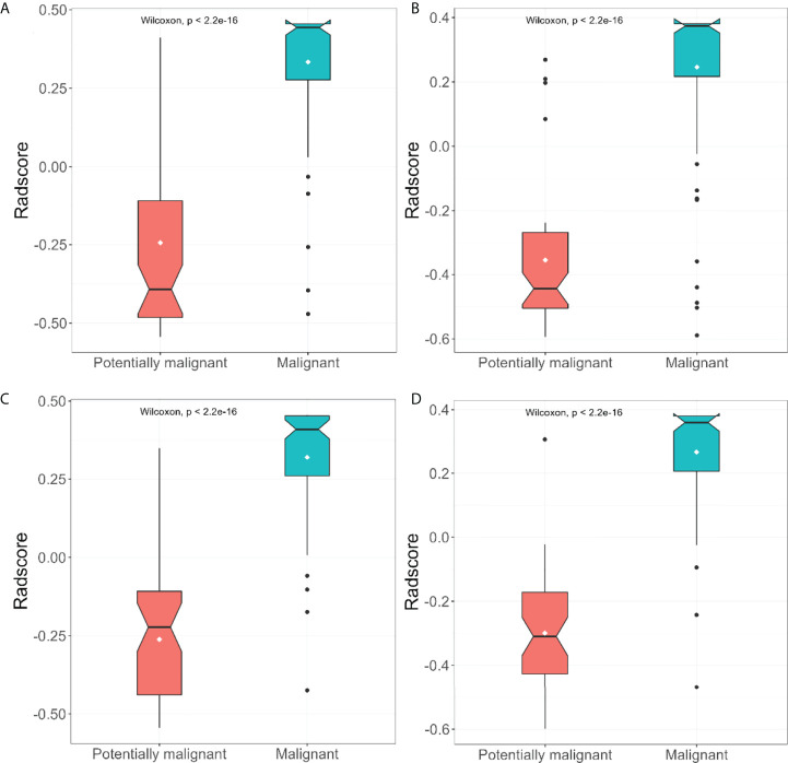
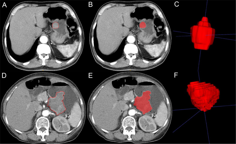
A total of 342 patients with GISTs confirmed histopathologically were enrolled from five medical centers. Data of patients wrom two centers comprised the training group (n=196), and data from the remaining three centers constituted the validation group (n=146). After CT image segmentation and feature extraction and selection, the arterial phase model and venous phase model were established. The maximum diameter of the tumor and internal necrosis were used to establish a clinical indicators model. The traditional CT diagnostic criteria were established for the classification of malignant potential of tumor. The performance of the four models was assessed using the receiver operating characteristics curve.

REUSLTS

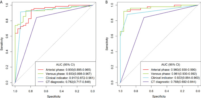
In the training group, the area under the curves(AUCs) of the arterial phase model, venous phase model, clinical indicators model, and traditional CT diagnostic criteria were 0.930 [95% confidence interval (CI): 0.895-0.965), 0.933 (95%CI 0.898-0.967), 0.917 (95%CI 0.872-0.961) and 0.782 (95%CI 0.717-0.848), respectively. In the validation group, the AUCs of the models were 0.960 (95%CI 0.930-0.990), 0.961 (95% CI 0.930-0.992), 0.922 (95%CI 0.884-0.960) and 0.768 (95%CI 0.692-0.844), respectively. No significant difference was detected in the AUC between the arterial phase model, venous phase model, and clinical indicators model by the DeLong test, whereas a significant difference was observed between the traditional CT diagnostic criteria and the other three models.

CONCLUSION

The radiomics model using the morphological features of GISTs play a significant role in tumor risk stratification and can provide a reference for clinical diagnosis and treatment plan.

摘要

目的

本研究旨在探讨基于增强CT图像的放射组学模型在鉴别胃肠道间质瘤(GIST)恶性风险方面的诊断效能,并与临床指标模型和传统CT诊断标准进行比较。

材料与方法

从五个医疗中心纳入342例经组织病理学确诊的GIST患者。来自两个中心的患者数据组成训练组(n = 196),其余三个中心的数据构成验证组(n = 146)。经过CT图像分割、特征提取与选择后,建立动脉期模型和静脉期模型。利用肿瘤最大直径和内部坏死情况建立临床指标模型。建立传统CT诊断标准用于肿瘤恶性潜能分类。采用受试者操作特征曲线评估四种模型的性能。

结果

在训练组中,动脉期模型、静脉期模型、临床指标模型和传统CT诊断标准的曲线下面积(AUC)分别为0.930 [95%置信区间(CI):0.895 - 0.965]、0.933(95%CI 0.898 - 0.967)、0.917(95%CI 0.872 - 0.961)和0.782(95%CI 0.717 - 0.848)。在验证组中,各模型的AUC分别为0.960(95%CI 0.930 - 0.990)、0.961(95%CI 0.930 - 0.992)、0.922(95%CI 0.884 - 0.960)和0.768(95%CI 0.692 - 0.844)。通过DeLong检验,动脉期模型、静脉期模型和临床指标模型的AUC之间未检测到显著差异,而传统CT诊断标准与其他三种模型之间存在显著差异。

结论

利用GIST形态学特征的放射组学模型在肿瘤风险分层中发挥重要作用,可为临床诊断和治疗方案提供参考。

https://cdn.ncbi.nlm.nih.gov/pmc/blobs/5c9b/9425090/ad356eab63c0/fonc-12-966743-g005.jpg
https://cdn.ncbi.nlm.nih.gov/pmc/blobs/5c9b/9425090/2376ef194147/fonc-12-966743-g001.jpg
https://cdn.ncbi.nlm.nih.gov/pmc/blobs/5c9b/9425090/009f41d14e0c/fonc-12-966743-g002.jpg
https://cdn.ncbi.nlm.nih.gov/pmc/blobs/5c9b/9425090/315635459536/fonc-12-966743-g003.jpg
https://cdn.ncbi.nlm.nih.gov/pmc/blobs/5c9b/9425090/412f68f5f058/fonc-12-966743-g004.jpg
https://cdn.ncbi.nlm.nih.gov/pmc/blobs/5c9b/9425090/ad356eab63c0/fonc-12-966743-g005.jpg

相似文献

1
Malignancy risk of gastrointestinal stromal tumors evaluated with noninvasive radiomics: A multi-center study.基于非侵入性影像组学评估胃肠道间质瘤的恶性风险:一项多中心研究
Front Oncol. 2022 Aug 16;12:966743. doi: 10.3389/fonc.2022.966743. eCollection 2022.
2
Value of contrast-enhanced CT based radiomic machine learning algorithm in differentiating gastrointestinal stromal tumors with KIT exon 11 mutation: a two-center study.基于增强 CT 的影像组学机器学习算法在鉴别 KIT 外显子 11 突变的胃肠道间质瘤中的价值:一项多中心研究。
Diagn Interv Radiol. 2022 Jan;28(1):29-38. doi: 10.5152/dir.2021.21600.
3
Radiomics Nomogram Based on Contrast-enhanced CT to Predict the Malignant Potential of Gastrointestinal Stromal Tumor: A Two-center Study.基于对比增强CT的影像组学列线图预测胃肠道间质瘤的恶性潜能:一项双中心研究
Acad Radiol. 2022 Jun;29(6):806-816. doi: 10.1016/j.acra.2021.05.005. Epub 2021 Jul 5.
4
Computed tomography-based radiomics model for discriminating the risk stratification of gastrointestinal stromal tumors.基于计算机断层扫描的放射组学模型用于鉴别胃肠道间质瘤的风险分层。
Radiol Med. 2020 May;125(5):465-473. doi: 10.1007/s11547-020-01138-6. Epub 2020 Feb 11.
5
Development of a multi-phase CT-based radiomics model to differentiate heterotopic pancreas from gastrointestinal stromal tumor.基于多期 CT 的放射组学模型的开发,用于鉴别异位胰腺与胃肠道间质瘤。
BMC Med Imaging. 2024 Feb 14;24(1):44. doi: 10.1186/s12880-024-01219-2.
6
Comparison of malignancy-prediction efficiency between contrast and non-contract CT-based radiomics features in gastrointestinal stromal tumors: A multicenter study.基于对比增强CT与非对比增强CT的影像组学特征在胃肠道间质瘤中恶性预测效率的比较:一项多中心研究
Clin Transl Med. 2020 Jul;10(3):e291. doi: 10.1002/ctm2.91. Epub 2020 Jul 7.
7
CT Radiomics Model for Discriminating the Risk Stratification of Gastrointestinal Stromal Tumors: A Multi-Class Classification and Multi-Center Study.用于鉴别胃肠道间质瘤风险分层的CT影像组学模型:一项多类别分类和多中心研究
Front Oncol. 2021 Jun 8;11:654114. doi: 10.3389/fonc.2021.654114. eCollection 2021.
8
Clinical-radiomics-based treatment decision support for KIT Exon 11 deletion in gastrointestinal stromal tumors: a multi-institutional retrospective study.基于临床影像组学的胃肠道间质瘤KIT外显子11缺失治疗决策支持:一项多机构回顾性研究
Front Oncol. 2023 Aug 14;13:1193010. doi: 10.3389/fonc.2023.1193010. eCollection 2023.
9
Building Radiomics Models Based on Triple-Phase CT Images Combining Clinical Features for Discriminating the Risk Rating in Gastrointestinal Stromal Tumors.基于三相CT图像联合临床特征构建放射组学模型以鉴别胃肠道间质瘤的风险分级
Front Oncol. 2021 Dec 7;11:737302. doi: 10.3389/fonc.2021.737302. eCollection 2021.
10
CT Radiomics for the Preoperative Prediction of Ki67 Index in Gastrointestinal Stromal Tumors: A Multi-Center Study.CT影像组学用于术前预测胃肠道间质瘤中Ki67指数:一项多中心研究
Front Oncol. 2021 Sep 14;11:689136. doi: 10.3389/fonc.2021.689136. eCollection 2021.

引用本文的文献

1
The Many Faces of Intestinal Tumors in Adults, Including the Primary Role of CT Imaging in Emergencies and the Important Role of Cross-Sectional Imaging: A Pictorial Review.成人肠道肿瘤的多种表现,包括CT成像在急诊中的主要作用及横断面成像的重要作用:图文综述
Healthcare (Basel). 2025 May 6;13(9):1071. doi: 10.3390/healthcare13091071.
2
Computed tomography radiomics models of tumor differentiation in canine small intestinal tumors.犬小肠肿瘤中肿瘤分化的计算机断层扫描放射组学模型
Front Vet Sci. 2024 Sep 23;11:1450304. doi: 10.3389/fvets.2024.1450304. eCollection 2024.
3
Advancing gastrointestinal stromal tumor management: The role of imagomics features in precision risk assessment.

本文引用的文献

1
TN-USMA Net: Triple normalization-based gastrointestinal stromal tumors classification on multicenter EUS images with ultrasound-specific pretraining and meta attention.TN-USMA 网络:基于三重归一化的多中心 EUS 图像胃间质瘤分类,采用超声特异性预训练和元注意力。
Med Phys. 2021 Nov;48(11):7199-7214. doi: 10.1002/mp.15172. Epub 2021 Aug 30.
2
Radiomics Nomogram Based on Contrast-enhanced CT to Predict the Malignant Potential of Gastrointestinal Stromal Tumor: A Two-center Study.基于对比增强CT的影像组学列线图预测胃肠道间质瘤的恶性潜能:一项双中心研究
Acad Radiol. 2022 Jun;29(6):806-816. doi: 10.1016/j.acra.2021.05.005. Epub 2021 Jul 5.
3
推进胃肠道间质瘤的管理:影像组学特征在精准风险评估中的作用。
World J Gastrointest Surg. 2024 Sep 27;16(9):2942-2952. doi: 10.4240/wjgs.v16.i9.2942.
4
Prediction of High-Risk Gastrointestinal Stromal Tumor Recurrence Based on Delta-CT Radiomics Modeling: A 3-Year Follow-up Study After Surgery.基于Delta-CT影像组学模型预测高危胃肠道间质瘤复发:术后3年随访研究
Clin Med Insights Oncol. 2024 Apr 15;18:11795549241245698. doi: 10.1177/11795549241245698. eCollection 2024.
5
Recurrent Gastrointestinal Stromal Tumor With Chondroid Differentiation After Imatinib Therapy: An Unusual Case and Literature Review.伊马替尼治疗后出现软骨样分化的复发性胃肠道间质瘤:1例罕见病例及文献复习
Cureus. 2024 Feb 24;16(2):e54842. doi: 10.7759/cureus.54842. eCollection 2024 Feb.
6
Relationship between multi-slice computed tomography features and pathological risk stratification assessment in gastric gastrointestinal stromal tumors.多层螺旋CT特征与胃胃肠道间质瘤病理风险分层评估之间的关系
World J Gastrointest Oncol. 2023 Jun 15;15(6):1073-1085. doi: 10.4251/wjgo.v15.i6.1073.
7
Development and Validation of Artificial-Intelligence-Based Radiomics Model Using Computed Tomography Features for Preoperative Risk Stratification of Gastrointestinal Stromal Tumors.基于计算机断层扫描特征的人工智能放射组学模型在胃肠道间质瘤术前风险分层中的开发与验证
J Pers Med. 2023 Apr 24;13(5):717. doi: 10.3390/jpm13050717.
8
Role of Machine Learning in Precision Oncology: Applications in Gastrointestinal Cancers.机器学习在精准肿瘤学中的作用:在胃肠道癌症中的应用
Cancers (Basel). 2022 Dec 22;15(1):63. doi: 10.3390/cancers15010063.
CT Radiomics Model for Discriminating the Risk Stratification of Gastrointestinal Stromal Tumors: A Multi-Class Classification and Multi-Center Study.
用于鉴别胃肠道间质瘤风险分层的CT影像组学模型:一项多类别分类和多中心研究
Front Oncol. 2021 Jun 8;11:654114. doi: 10.3389/fonc.2021.654114. eCollection 2021.
4
Value of radiomics model based on enhanced computed tomography in risk grade prediction of gastrointestinal stromal tumors.基于增强 CT 的影像组学模型预测胃肠道间质瘤危险度分级的价值
Sci Rep. 2021 Jun 8;11(1):12009. doi: 10.1038/s41598-021-91508-5.
5
MRI-Based Radiomics Models for Predicting Risk Classification of Gastrointestinal Stromal Tumors.基于MRI的放射组学模型预测胃肠道间质瘤的风险分类
Front Oncol. 2021 May 10;11:631927. doi: 10.3389/fonc.2021.631927. eCollection 2021.
6
Computed-Tomography-Based Radiomics Model for Predicting the Malignant Potential of Gastrointestinal Stromal Tumors Preoperatively: A Multi-Classifier and Multicenter Study.基于计算机断层扫描的放射组学模型术前预测胃肠道间质瘤恶性潜能:多分类器和多中心研究
Front Oncol. 2021 Apr 22;11:582847. doi: 10.3389/fonc.2021.582847. eCollection 2021.
7
Assessment of morphological CT imaging features for the prediction of risk stratification, mutations, and prognosis of gastrointestinal stromal tumors.评估形态 CT 成像特征对胃肠道间质瘤风险分层、突变和预后的预测。
Eur Radiol. 2021 Nov;31(11):8554-8564. doi: 10.1007/s00330-021-07961-3. Epub 2021 Apr 21.
8
Exploring the predictive value of additional peritumoral regions based on deep learning and radiomics: A multicenter study.基于深度学习和放射组学的瘤周额外区域预测价值的探索:一项多中心研究。
Med Phys. 2021 May;48(5):2374-2385. doi: 10.1002/mp.14767. Epub 2021 Mar 30.
9
MRI Texture-Based Models for Predicting Mitotic Index and Risk Classification of Gastrointestinal Stromal Tumors.MRI 纹理模型预测胃肠道间质瘤有丝分裂指数和危险度分级。
J Magn Reson Imaging. 2021 Apr;53(4):1054-1065. doi: 10.1002/jmri.27390. Epub 2020 Oct 9.
10
New advances in radiomics of gastrointestinal stromal tumors.胃肠道间质瘤的放射组学新进展
World J Gastroenterol. 2020 Aug 28;26(32):4729-4738. doi: 10.3748/wjg.v26.i32.4729.