Chen Zhonghua, Xu Linyi, Zhang Chuanmin, Huang Chencui, Wang Minhong, Feng Zhan, Xiong Yue
Department of Radiology, Haining People's Hospital, Jiaxing, China.
Department of Research Collaboration, R&D Center, Beijing Deepwise & League of PHD Technology Co., Ltd, R&D Center, Beijing, China.
Front Oncol. 2021 Jun 8;11:654114. doi: 10.3389/fonc.2021.654114. eCollection 2021.
To establish and verify a computed tomography (CT)-based multi-class prediction model for discriminating the risk stratification of gastrointestinal stromal tumors (GISTs).
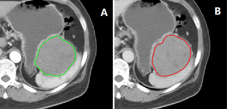
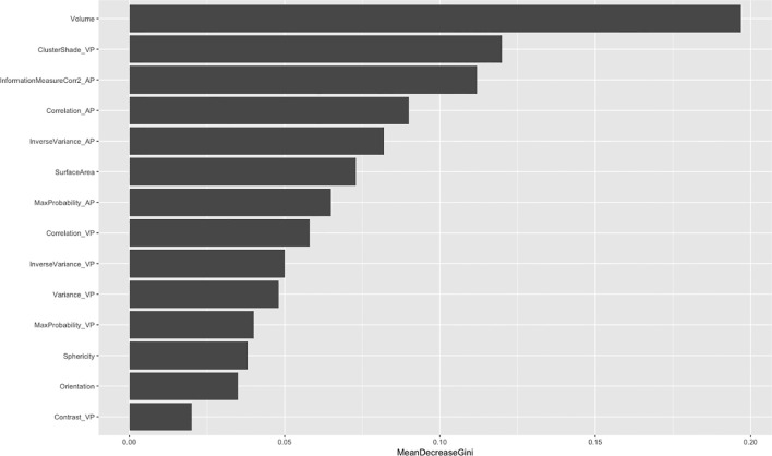
A total of 381 patients with GISTs were confirmed by surgery and pathology. Information on 213 patients were obtained from one hospital and used as training cohort, whereas the details of 168 patients were collected from two other hospitals and used as independent validation cohort. Regions of interest on CT images of arterial and venous phases were drawn, radiomics features were extracted, and dimensionality reduction processing was performed. Using a one-vs-rest method, a Random Forest-based GISTs risk three-class prediction model was established, and the receiver operating characteristic curve (ROC) was used to evaluate the performance of the multi-class classification model, and the generalization ability was verified using external data.
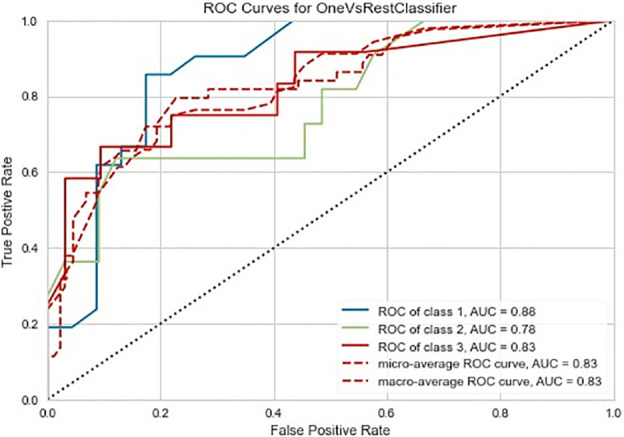
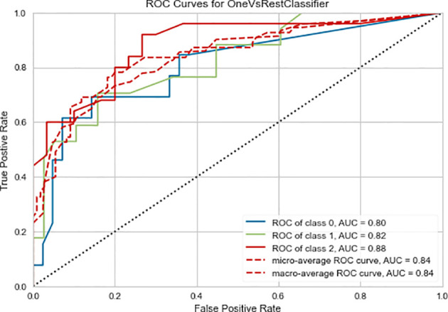
The training cohort included 96 very low-risk and low-risk, 60 intermediate-risk and 57 high-risk patients. External validation cohort included 82 very low-risk and low-risk, 48 intermediate-risk and 38 high-risk patients. The GISTs risk three-class radiomics model had a macro/micro average area under the curve (AUC) of 0.84 and an accuracy of 0.78 in the training cohort. It had a stable performance in the external validation cohort, with a macro/micro average AUC of 0.83 and an accuracy of 0.80.
CT radiomics can discriminate GISTs risk stratification. The performance of the three-class radiomics prediction model is good, and its generalization ability has also been verified in the external validation cohort, indicating its potential to assist stratified and accurate treatment of GISTs in the clinic.
建立并验证基于计算机断层扫描(CT)的多分类预测模型,以鉴别胃肠道间质瘤(GISTs)的风险分层。
共有381例GISTs患者经手术及病理确诊。其中213例患者的信息来自一家医院,用作训练队列,而另外168例患者的详细信息则从其他两家医院收集,用作独立验证队列。绘制动脉期和静脉期CT图像上的感兴趣区域,提取影像组学特征,并进行降维处理。采用一对多方法,建立基于随机森林的GISTs风险三分类预测模型,采用受试者操作特征曲线(ROC)评估多分类模型的性能,并使用外部数据验证其泛化能力。
训练队列包括96例极低风险和低风险、60例中度风险和57例高风险患者。外部验证队列包括82例极低风险和低风险、48例中度风险和38例高风险患者。GISTs风险三分类影像组学模型在训练队列中的曲线下宏/微平均面积(AUC)为0.84,准确率为0.78。其在外部验证队列中表现稳定,宏/微平均AUC为0.83,准确率为0.80。
CT影像组学可鉴别GISTs的风险分层。三分类影像组学预测模型性能良好,其泛化能力也在外部验证队列中得到验证,表明其在临床中辅助GISTs分层及精准治疗的潜力。