Department of Cardiology, State Key Laboratory of Organ Failure Research, Nanfang Hospital, Southern Medical University, 510515, Guangzhou, China; Guangdong Provincial Key Laboratory of Cardiac Function and Microcirculation, 510515, Guangzhou, China.
Department of Cardiology, State Key Laboratory of Organ Failure Research, Nanfang Hospital, Southern Medical University, 510515, Guangzhou, China; Guangdong Provincial Key Laboratory of Cardiac Function and Microcirculation, 510515, Guangzhou, China; Guizhou University Hospital, Guiyang Guizhou, 550025, China.
Redox Biol. 2022 Oct;56:102446. doi: 10.1016/j.redox.2022.102446. Epub 2022 Aug 23.
Metabolic switching during heart development contributes to postnatal cardiomyocyte (CM) cell cycle exit and loss of regenerative capacity in the mammalian heart. Metabolic control has potential for developing effective CM proliferation strategies. We sought to determine whether lactate dehydrogenase A (LDHA) regulated CM proliferation by inducing metabolic reprogramming.
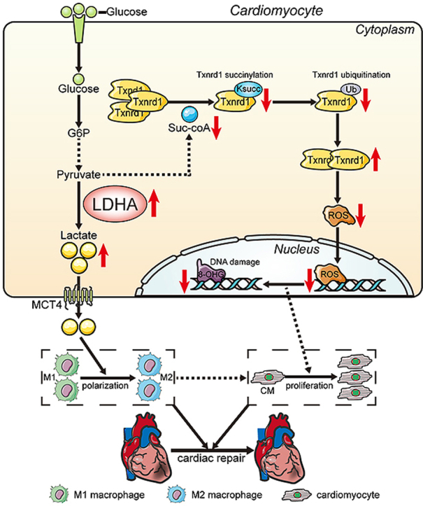
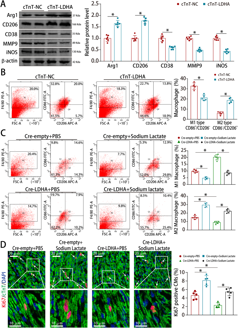
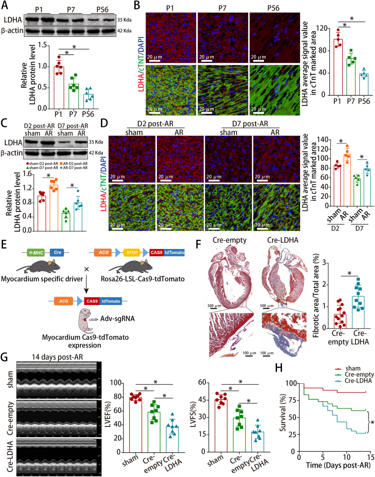
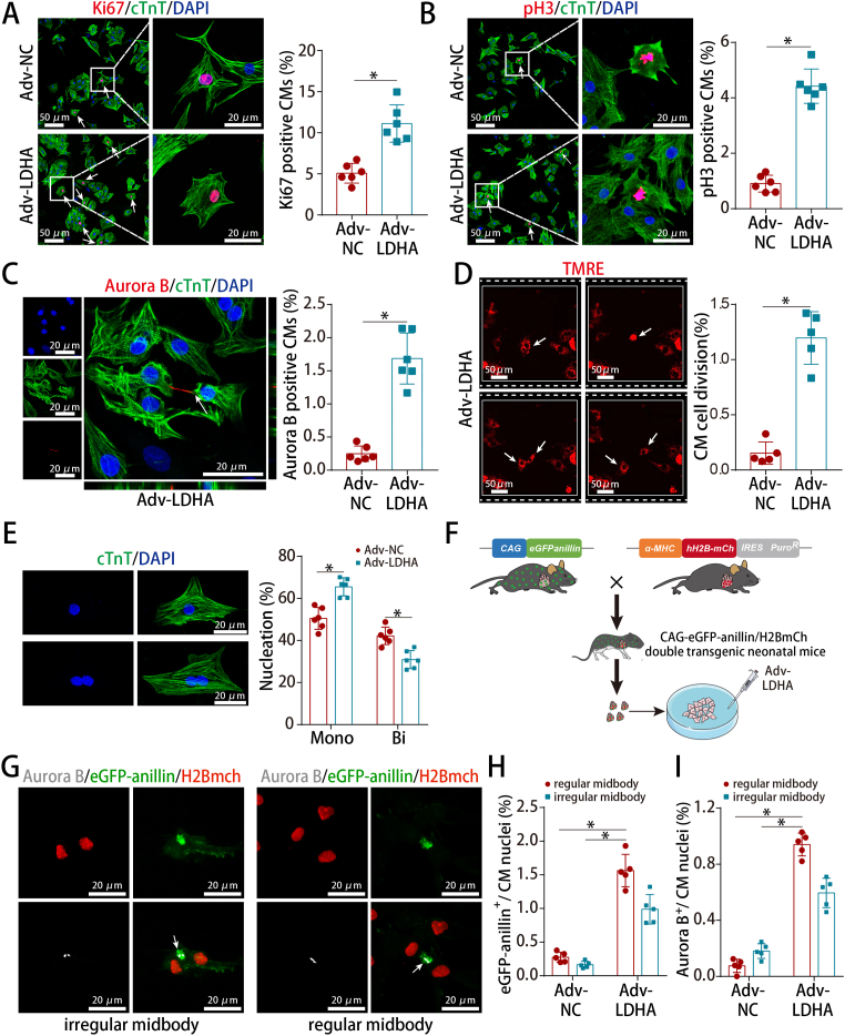
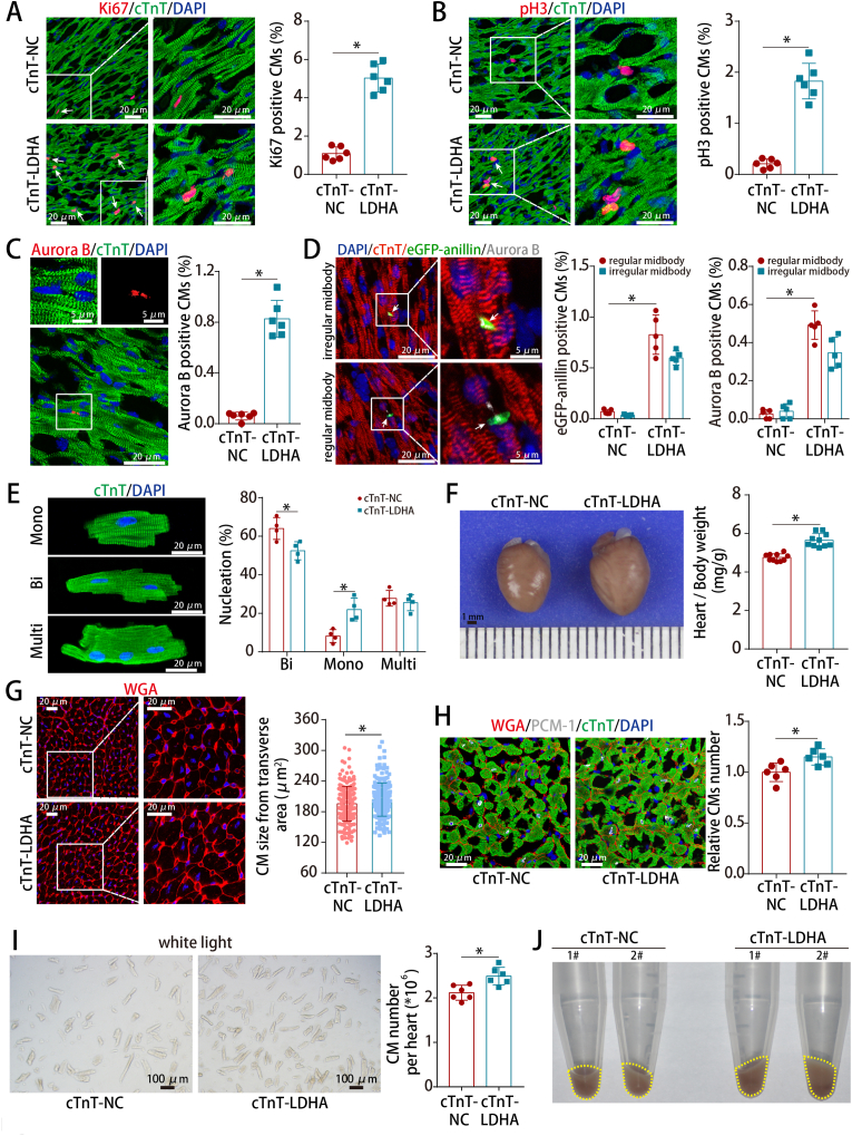
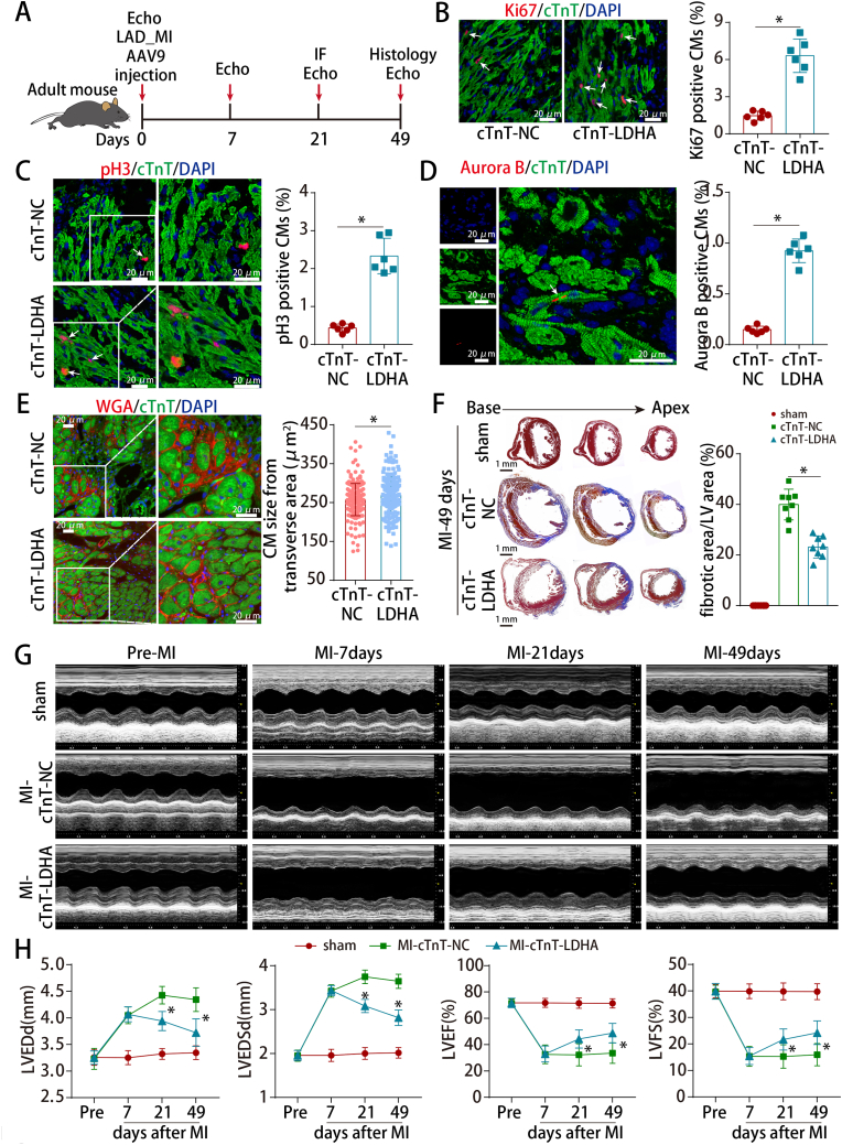
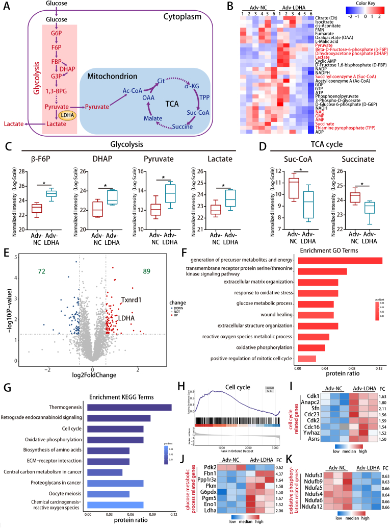
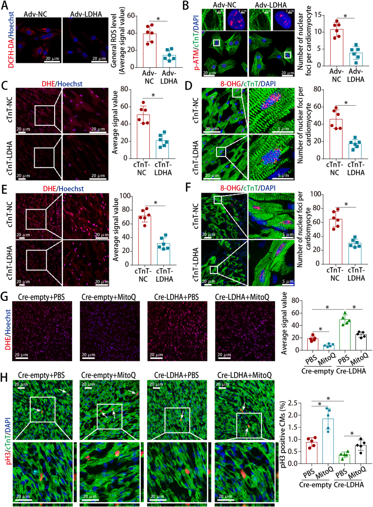
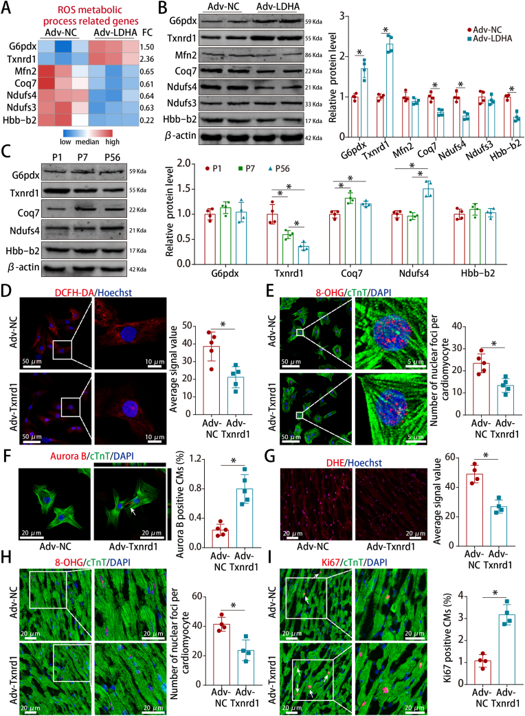
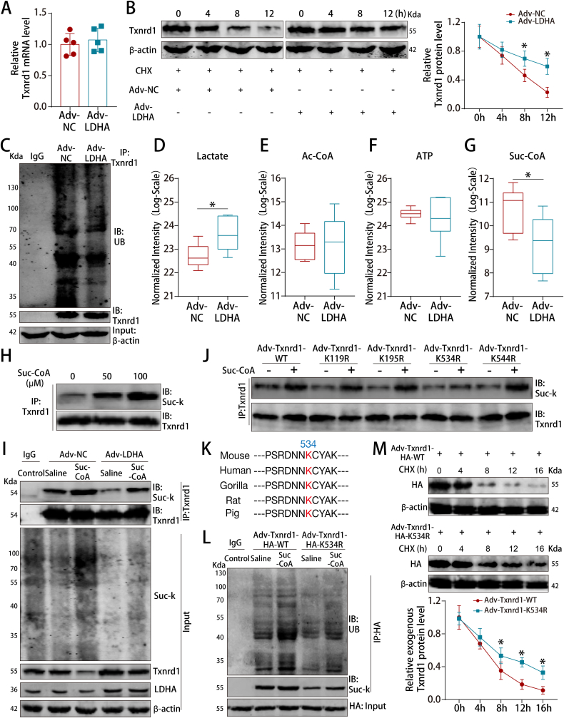
LDHA expression was high in P1 hearts and significantly decreased during postnatal heart development. CM-specific LDHA knockout mice were generated using CRISPR/Cas9 technology. CM-specific LDHA knockout inhibited CM proliferation, leading to worse cardiac function and a lower survival rate in the neonatal apical resection model. In contrast, CM-specific overexpression of LDHA promoted CM proliferation and cardiac repair post-MI. The α-MHC-H2B-mCh/CAG-eGFP-anillin system was used to confirm the proliferative effect triggered by LDHA on P7 CMs and adult hearts. Metabolomics, proteomics and Co-IP experiments indicated that LDHA-mediated succinyl coenzyme A reduction inhibited succinylation-dependent ubiquitination of thioredoxin reductase 1 (Txnrd1), which alleviated ROS and thereby promoted CM proliferation. In addition, flow cytometry and western blotting showed that LDHA-driven lactate production created a beneficial cardiac regenerative microenvironment by inducing M2 macrophage polarization.
LDHA-mediated metabolic reprogramming promoted CM proliferation by alleviating ROS and inducing M2 macrophage polarization, indicating that LDHA might be an effective target for promoting cardiac repair post-MI.
心脏发育过程中的代谢转换有助于哺乳动物心脏出生后心肌细胞 (CM) 停止细胞周期并丧失再生能力。代谢控制有可能开发出有效的 CM 增殖策略。我们试图确定 LDHA 是否通过诱导代谢重编程来调节 CM 增殖。
在 P1 心脏中 LDHA 表达水平较高,并在出生后心脏发育过程中显著降低。使用 CRISPR/Cas9 技术生成了 CM 特异性 LDHA 敲除小鼠。CM 特异性 LDHA 敲除抑制 CM 增殖,导致新生儿心尖切除模型中心脏功能恶化和存活率降低。相比之下,CM 特异性 LDHA 过表达促进了 MI 后 CM 增殖和心脏修复。使用 α-MHC-H2B-mCh/CAG-eGFP-anillin 系统证实了 LDHA 对 P7 CM 和成年心脏触发的增殖作用。代谢组学、蛋白质组学和 Co-IP 实验表明,LDHA 介导的琥珀酰辅酶 A 还原抑制了硫氧还蛋白还原酶 1 (Txnrd1) 的琥珀酰化依赖性泛素化,从而减轻了 ROS,从而促进了 CM 增殖。此外,流式细胞术和 Western blot 表明,LDHA 驱动的乳酸产生通过诱导 M2 巨噬细胞极化创造了有益的心脏再生微环境。
LDHA 介导的代谢重编程通过减轻 ROS 和诱导 M2 巨噬细胞极化来促进 CM 增殖,表明 LDHA 可能是促进 MI 后心脏修复的有效靶点。