Department of Internal Medicine, Toho University Graduate School of Medicine, Tokyo, Japan.
Division of Rheumatology, Department of Internal Medicine, Toho University School of Medicine, Tokyo, Japan.
Front Immunol. 2022 Aug 18;13:935114. doi: 10.3389/fimmu.2022.935114. eCollection 2022.
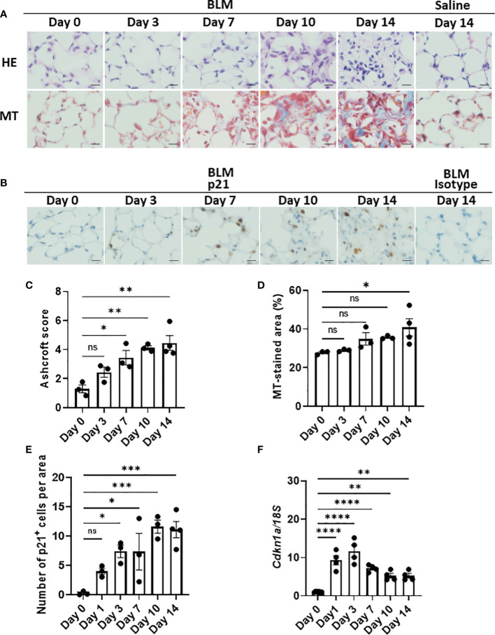
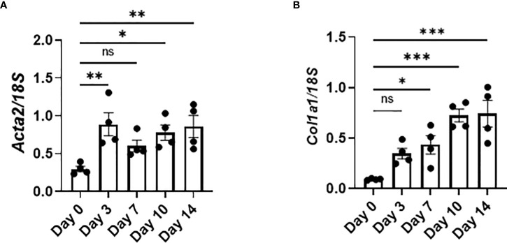
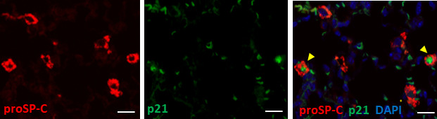
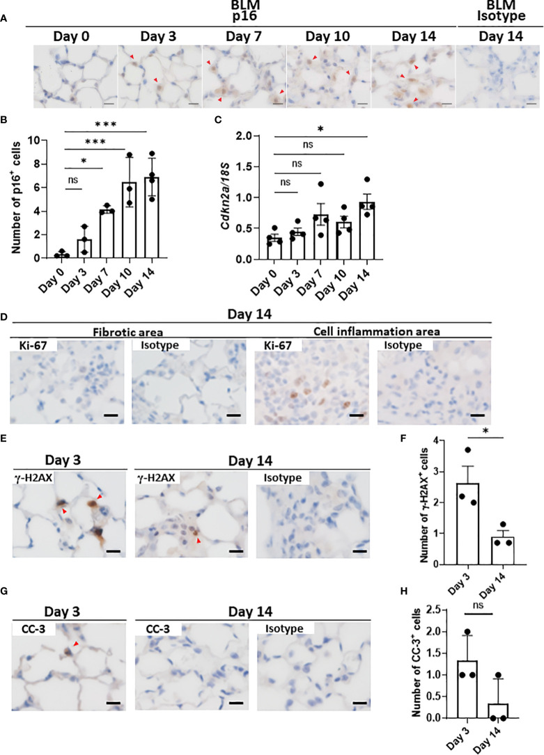
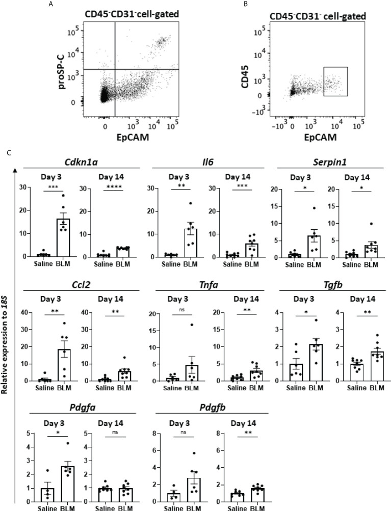
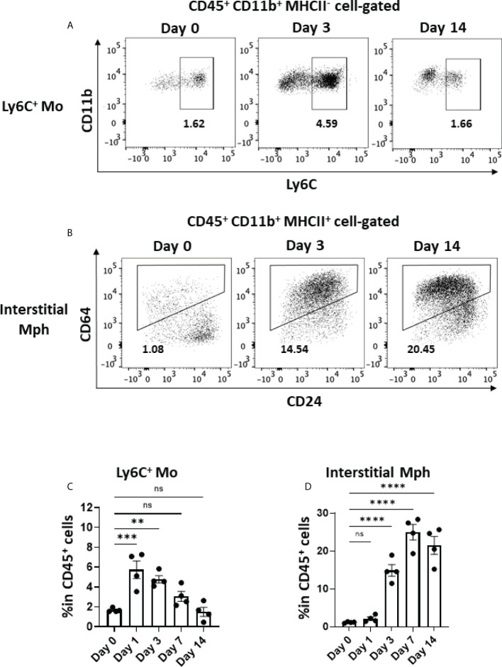
Fibrosing interstitial lung disease (ILD) develops due to the impaired reparative processes following lung tissue damage. Cellular senescence has been reported to contribute to the progression of fibrosis. However, the mechanisms by which these senescent cells initiate and/or drive the progression of lung tissue fibrosis are not yet fully understood. We demonstrated that p21- and p16-pathway-dependent senescence in type 2 alveolar epithelial cells (AEC2) were both involved in the initiation and progression of lung fibrosis in murine bleomycin (BLM)-induced ILD. p21-senescent AEC2 emerged rapidly, as early as 1 day after the intratracheal instillation of BLM. Their number subsequently increased and persisted until the later fibrosis phase. Very few p16-senescent AEC2 emerged upon the instillation of BLM, and their increase was slower and milder than that of p21 AEC2. AEC2 enriched with senescent cells sorted from BLM-ILD lungs expressed senescence-associated secretory phenotype (SASP)-related genes, including , , , , , and , at the initiation and chronic phases of fibrosis, exhibiting distinct expression patterns of magnitude that were dependent on the disease phase. Ly6C inflammatory monocytes increased in the lungs immediately after the instillation of BLM and interstitial macrophages increased from day 3. The expression of and was upregulated as early as day 1, indicating the activation of fibroblasts. We speculated that IL-6, plasminogen activator inhibitor-1 (PAI-1), and TGF-β contributed to the accumulation of senescent cells during the progression of fibrosis in an autocrine and paracrine manner. In addition, CCL2, produced in large amounts by senescent AEC2, may have induced the infiltration of Ly6C inflammatory monocytes in the early phase, and TGF-β and PDGFa from senescent AEC2 may contribute to the activation of fibroblasts in the very early phases. Our study indicated that senescent AEC2 plays a role in the pathogenesis of fibrosing ILD throughout the course of the disease and provides insights into its pathogenesis, which may lead to the development of new therapeutic methods targeting senescent cells or SASP molecules.
纤维性间质性肺病(ILD)是由于肺组织损伤后的修复过程受损而发展起来的。细胞衰老已被报道有助于纤维化的进展。然而,这些衰老细胞引发和/或驱动肺组织纤维化进展的机制尚不完全清楚。我们证明,2 型肺泡上皮细胞(AEC2)中 p21 和 p16 通路依赖性衰老均参与了博莱霉素(BLM)诱导的 ILD 中肺纤维化的起始和进展。p21 衰老的 AEC2 迅速出现,早在 BLM 气管内滴注后 1 天即可出现。它们的数量随后增加并持续到后期纤维化阶段。BLM 滴注后很少出现 p16 衰老的 AEC2,其增加速度比 p21 AEC2 慢且温和。从 BLM-ILD 肺中分离出来的富含衰老细胞的 AEC2 表达了衰老相关分泌表型(SASP)相关基因,包括、、、、、和,在纤维化的起始和慢性阶段,表现出依赖于疾病阶段的不同表达模式。BLM 滴注后,Ly6C 炎症单核细胞立即在肺部增加,间质巨噬细胞从第 3 天开始增加。第 1 天就上调了和的表达,表明成纤维细胞被激活。我们推测,IL-6、纤溶酶原激活物抑制剂-1(PAI-1)和 TGF-β 通过自分泌和旁分泌方式促进纤维化进展过程中衰老细胞的积累。此外,大量由衰老 AEC2 产生的 CCL2 可能在早期诱导了 Ly6C 炎症单核细胞的浸润,而来自衰老 AEC2 的 TGF-β 和 PDGFa 可能有助于成纤维细胞在极早期的激活。我们的研究表明,衰老的 AEC2 在整个疾病过程中在纤维性间质性肺病的发病机制中发挥作用,并深入了解其发病机制,这可能导致针对衰老细胞或 SASP 分子的新治疗方法的发展。