Guangdong Provincial Geriatrics Institute, Guangdong Provincial People's Hospital, Guangdong Academy of Medical Sciences, Guangzhou 510080, China.
Department of Medicine, Brigham and Women's Hospital and Harvard Medical School, Boston, MA 02115, USA.
Cardiovasc Res. 2023 May 2;119(4):1046-1061. doi: 10.1093/cvr/cvac144.
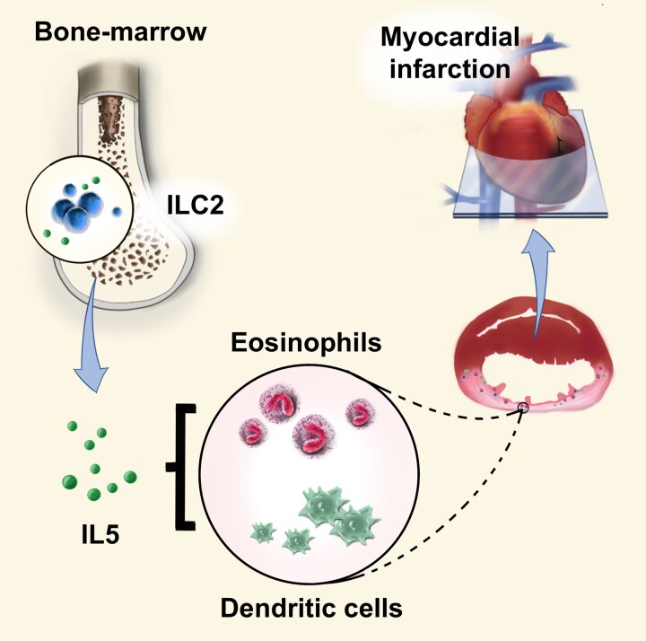
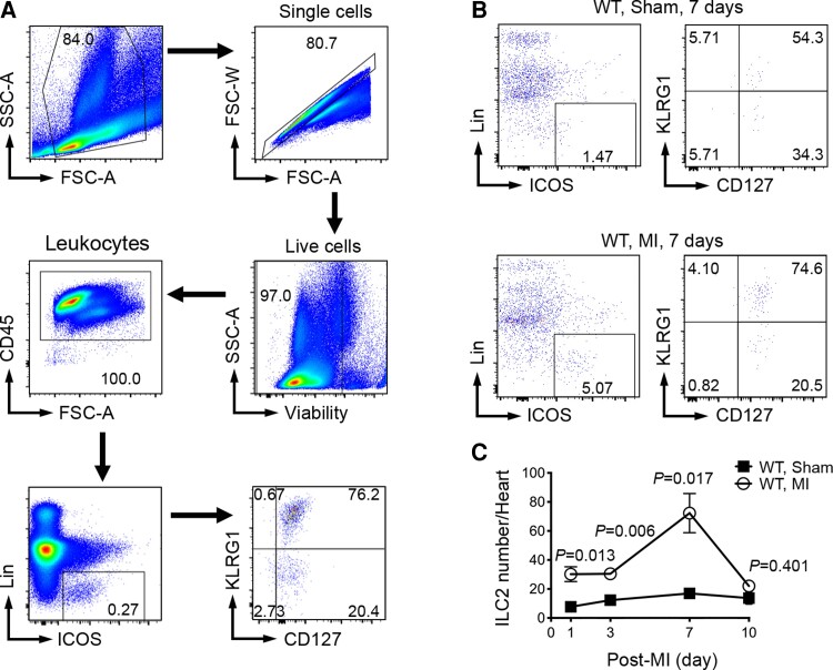
Group 2 innate lymphoid cells (ILC2s) regulate adaptive and innate immunities. In mouse heart, production of myocardial infarction (MI) increased ILC2 accumulation, suggesting a role for ILC2 in cardiac dysfunction post-MI.
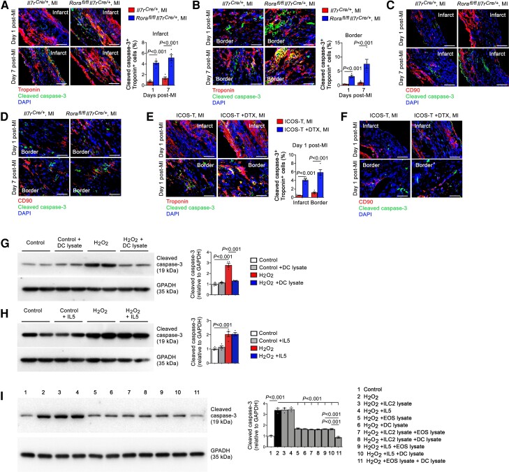
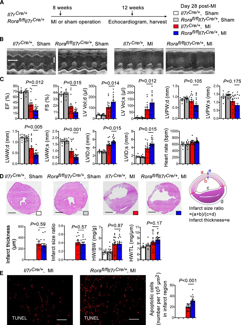
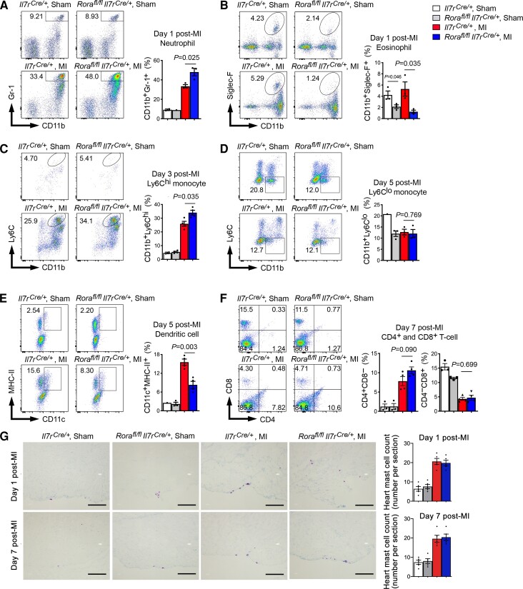
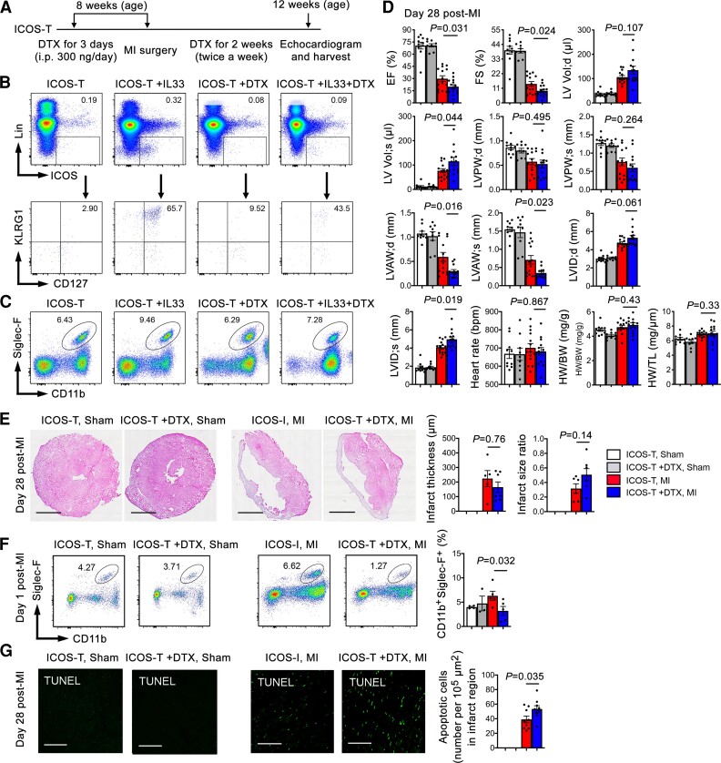
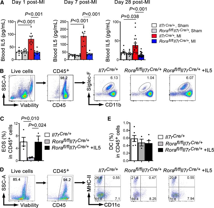
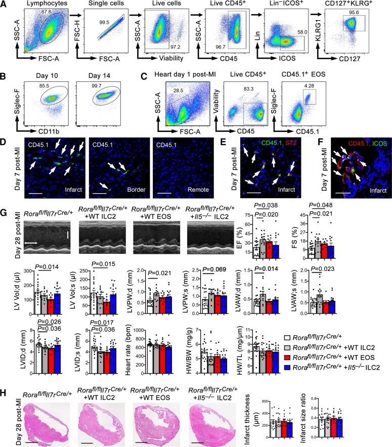
We produced MI in ILC2-deficeint Rorafl/flIl7rCre/+ mice and in Icosfl-DTR-fl/+Cd4Cre/+ mice that allowed diphtheria toxin-induced ILC2 depletion. Genetic or induced deficiency of ILC2 in mice exacerbated cardiac dysfunction post-MI injury along with increased myocardial accumulation of neutrophils, CD11b+Ly6Chi monocytes, and CD4+ T cells but deficiency of eosinophils (EOS) and dendritic cells (DC). Post-MI hearts from genetic and induced ILC2-deficient mice contained many more apoptotic cells than those of control mice, and Rorafl/flIl7rCre/+ mice showed thinner and larger infarcts and more collagen-I depositions than the Il7rCre/+ mice only at early time points post-MI. Mechanistic studies revealed elevated blood IL5 in Il7rCre/+ mice at 1, 7, and 28 days post-MI. Such increase was blunted in Rorafl/flIl7rCre/+ mice. Administration of recombinant IL5 reversed EOS losses in Rorafl/flIl7rCre/+ mice, but IL5 did not correct the DC loss in these mice. Adoptive transfer of ILC2, EOS, or DC from wild-type mice, but not ILC2 from Il5-/- mice improved post-MI cardiac functions in Rorafl/flIl7rCre/+ recipient mice. EOS are known to protect cardiomyocytes from apoptosis. Here we showed that DC acted like EOS in blocking cardiomyocyte apoptosis. Yet, ILC2 or IL5 alone did not directly affect cardiomyocyte apoptosis or TGF-β (transforming growth factor-β)-induced cardiac fibroblast Smad signalling.
This study revealed an indirect cardiac reparative role of ILC2 in post-MI hearts via the IL5, EOS, and DC mechanism.
2 型固有淋巴细胞 (ILC2) 调节适应性和固有免疫。在小鼠心脏中,心肌梗死 (MI) 增加了 ILC2 的积累,表明 ILC2 在 MI 后心脏功能障碍中起作用。
我们在 ILC2 缺陷型 Rorafl/flIl7rCre/+ 小鼠和 Icosfl-DTR-fl/+Cd4Cre/+ 小鼠中产生 MI,这些小鼠允许使用白喉毒素诱导 ILC2 耗竭。在 MI 损伤后,小鼠中 ILC2 的遗传或诱导缺失加剧了心脏功能障碍,同时伴有中性粒细胞、CD11b+Ly6Chi 单核细胞和 CD4+T 细胞的心肌积累增加,但嗜酸性粒细胞 (EOS) 和树突状细胞 (DC) 缺失。与对照小鼠相比,遗传和诱导的 ILC2 缺陷小鼠的 MI 后心脏含有更多的凋亡细胞,并且 Rorafl/flIl7rCre/+ 小鼠在 MI 后早期比 Il7rCre/+ 小鼠表现出更薄和更大的梗死面积和更多的胶原-I 沉积。机制研究显示,在 MI 后 1、7 和 28 天,Il7rCre/+ 小鼠的血液中 IL5 升高。这种增加在 Rorafl/flIl7rCre/+ 小鼠中减弱。重组 IL5 的给药逆转了 Rorafl/flIl7rCre/+ 小鼠中的 EOS 丢失,但在这些小鼠中,IL5 并未纠正 DC 丢失。从野生型小鼠中过继转移 ILC2、EOS 或 DC,但从 Il5-/- 小鼠中过继转移 ILC2 可改善 Rorafl/flIl7rCre/+ 受体小鼠的 MI 后心脏功能。已知 EOS 可保护心肌细胞免于凋亡。在这里,我们表明 DC 类似于 EOS,可阻止心肌细胞凋亡。然而,ILC2 或 IL5 单独不能直接影响心肌细胞凋亡或 TGF-β(转化生长因子-β)诱导的心肌成纤维细胞 Smad 信号传导。
这项研究揭示了 ILC2 通过 IL5、EOS 和 DC 机制在 MI 后心脏中的间接心脏修复作用。