Department of Esthetic and Restorative Dentistry, Saint Joseph University of Beirut, Lebanon.
Department of Neurosciences, Reproductive and Odontostomatological Sciences, University of Naples "Federico II", 80131 Napoli, Italy.
Biomed Res Int. 2022 Aug 27;2022:2805343. doi: 10.1155/2022/2805343. eCollection 2022.
This study is aimed at determining two main points. First, if the Canary System™ (CS), initially used to assess caries, can measure a decalcification depth of bleached enamel quantitatively, and second, whether or not whitening has a harmful effect on enamel. This device can be considered a useful tool in the clinical assessment of the progression of demineralization after bleaching.
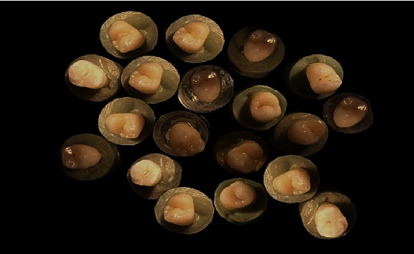
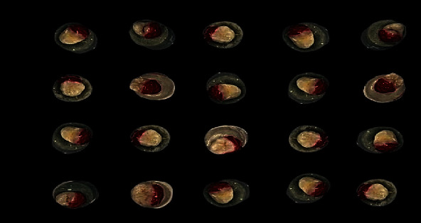
This study collected sixty human premolars that are in a good state recently extracted for orthodontic reason. To properly disinfect and preserve the premolars, they were stored in a saline solution and later in distilled water for a period of two weeks to allow the premolars to rehydrate. Later, 24 hours before the experiment, the premolars were introduced into a solution of artificial saliva to acquire back their minerals. The mineral content of the teeth was measured by the Canary System™ before bleaching. The teeth were bleached with 30% hydrogen peroxide (fläsh HP 30%), 30 min per week and for 3 consecutive weeks to simulate the conditions of strong bleaching in the clinic. The extent of demineralized enamel was measured by the Canary System™ at three points on the enamel surface of each tooth. The data were averaged for each application of the bleaching product. The demineralization extent of the teeth was measured by the Canary System™ before and after bleaching. The significance level was set at 0.05, and SPSS version 26 was used. The data were analyzed by using Wilcoxon's and Student's tests.
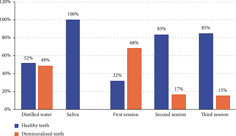
Mineral loss occurred after the first bleaching session; the Canary System™ detected a decalcification in the first bleaching session (532 ± 322 m) compared to the other sessions ( ≤ 0.05), while no significant change was detected between the second and the third sessions ( > 0.05).
Based on the findings of the present study, under in vitro conditions, it was possible to measure the demineralization extent of bleached enamel with the Canary System™.
本研究旨在确定两点。首先,如果 Canary System™(CS)最初用于评估龋齿,是否可以定量测量漂白牙釉质的脱矿深度,其次,漂白是否对牙釉质有有害影响。该设备可以被认为是临床评估漂白后脱矿进展的有用工具。
本研究收集了六十颗最近因正畸原因而拔除的人类前磨牙。为了正确消毒和保存前磨牙,将其储存在盐溶液中,然后在蒸馏水中储存两周,以使前磨牙重新水化。之后,在实验前 24 小时,将前磨牙置于人工唾液溶液中以恢复其矿物质。在漂白之前,使用 Canary System™ 测量牙齿的矿物质含量。用 30%过氧化氢(fläsh HP 30%)每周漂白 30 分钟,连续 3 周,以模拟临床强漂白条件。使用 Canary System™ 在每个牙齿表面的三个点测量脱矿牙釉质的程度。每次应用漂白产品后对数据进行平均。用 Canary System™ 测量漂白前后牙齿的脱矿程度。显著性水平设定为 0.05,使用 SPSS 版本 26。使用 Wilcoxon 和 Student 检验分析数据。
第一次漂白后发生了矿物质损失;Canary System™ 在第一次漂白(532 ± 322 μm)中检测到脱矿,与其他次(≤0.05)相比,而在第二次和第三次之间没有检测到显著变化(>0.05)。
根据本研究的结果,在体外条件下,Canary System™ 可以测量漂白牙釉质的脱矿程度。