• 文献检索
  • 文档翻译
  • 深度研究
  • 学术资讯
  • Suppr Zotero 插件Zotero 插件
  • 邀请有礼
  • 套餐&价格
  • 历史记录
应用&插件
Suppr Zotero 插件Zotero 插件浏览器插件Mac 客户端Windows 客户端微信小程序
定价
高级版会员购买积分包购买API积分包
服务
文献检索文档翻译深度研究API 文档MCP 服务
关于我们
关于 Suppr公司介绍联系我们用户协议隐私条款
关注我们

Suppr 超能文献

核心技术专利:CN118964589B侵权必究
粤ICP备2023148730 号-1Suppr @ 2026

文献检索

告别复杂PubMed语法,用中文像聊天一样搜索,搜遍4000万医学文献。AI智能推荐,让科研检索更轻松。

立即免费搜索

文件翻译

保留排版,准确专业,支持PDF/Word/PPT等文件格式,支持 12+语言互译。

免费翻译文档

深度研究

AI帮你快速写综述,25分钟生成高质量综述,智能提取关键信息,辅助科研写作。

立即免费体验

瘦素通过脑内出血时的周细胞 STAT3 通路促进血管生成。

Leptin Promotes Angiogenesis via Pericyte STAT3 Pathway upon Intracerebral Hemorrhage.

机构信息

Laboratory of Neuroscience, Affiliated Hospital of Guilin Medical University, Guilin Medical University, Guilin 541004, China.

Department of Neurology, Affiliated Hospital of Guilin Medical University, Guilin Medical University, Guilin 541004, China.

出版信息

Cells. 2022 Sep 3;11(17):2755. doi: 10.3390/cells11172755.

DOI:10.3390/cells11172755
PMID:36078162
原文链接:https://pmc.ncbi.nlm.nih.gov/articles/PMC9454866/
Abstract

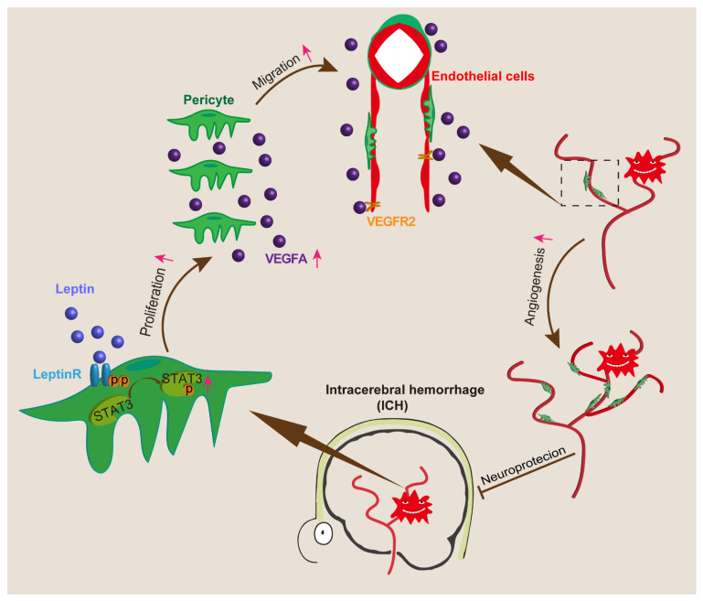
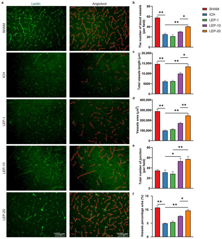
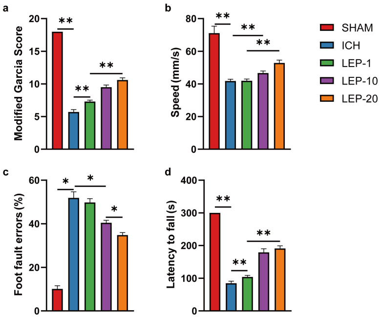
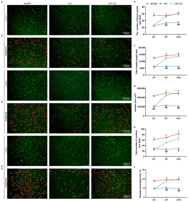
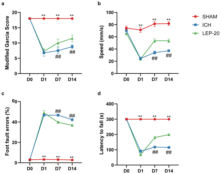
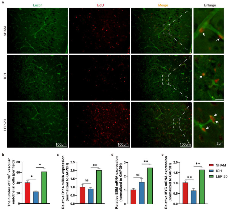
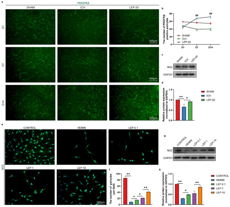
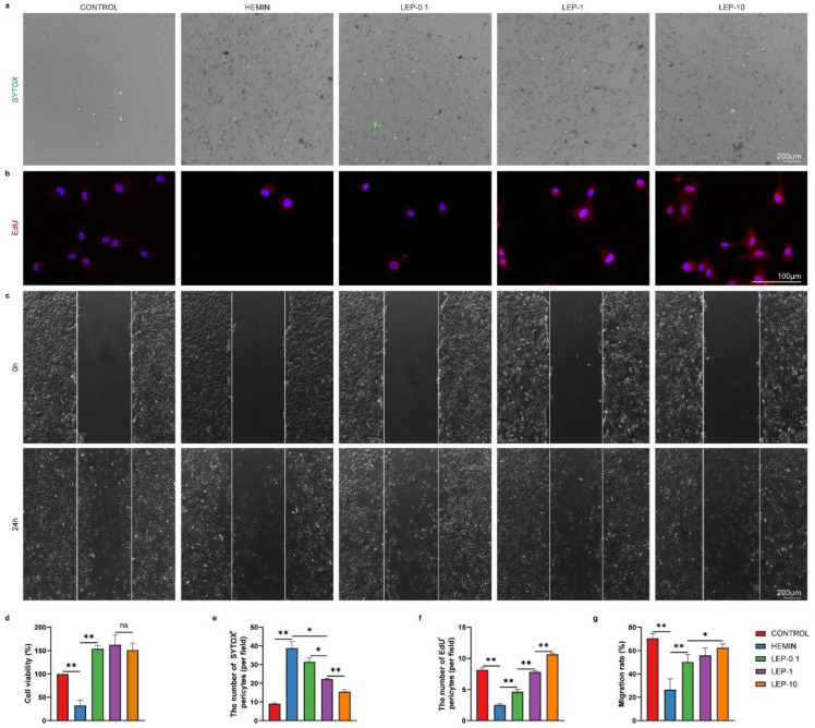
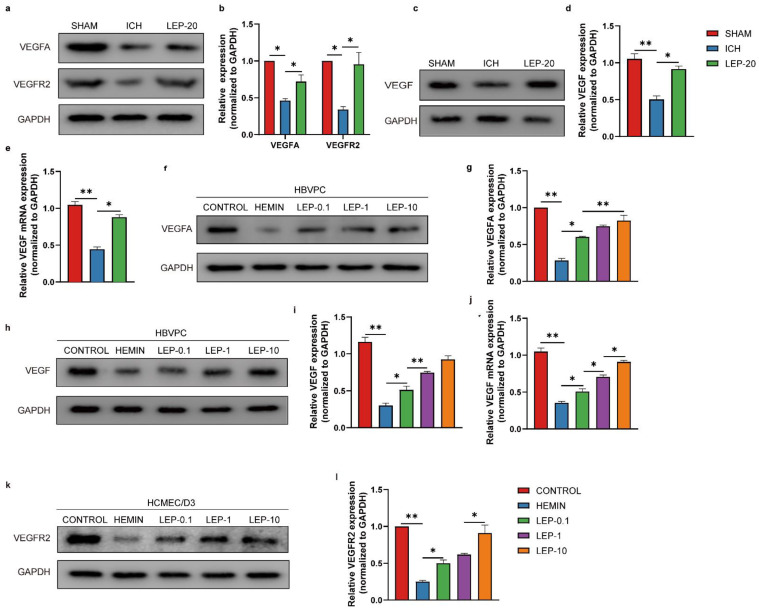
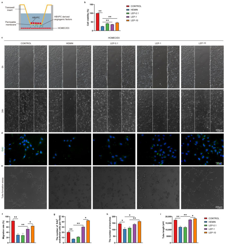
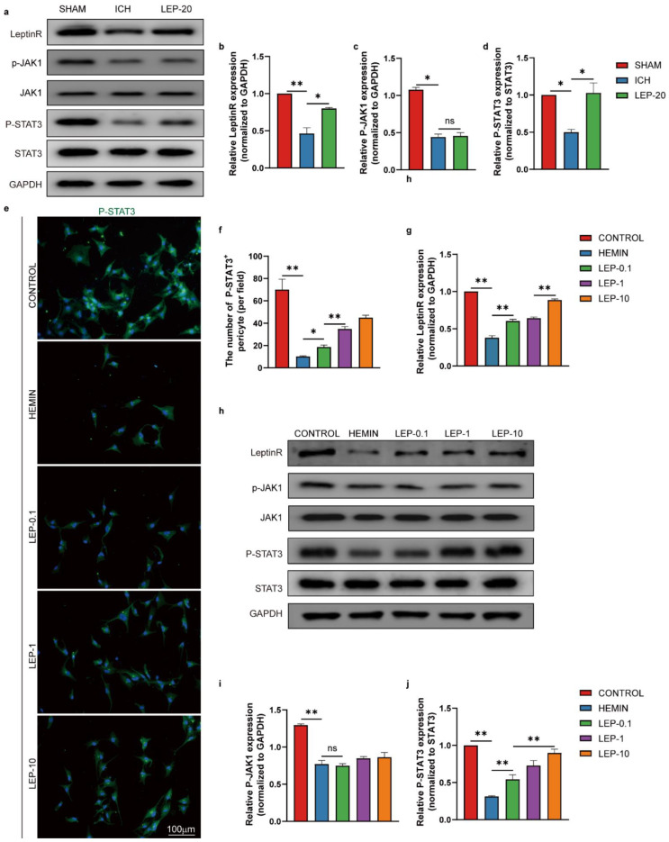
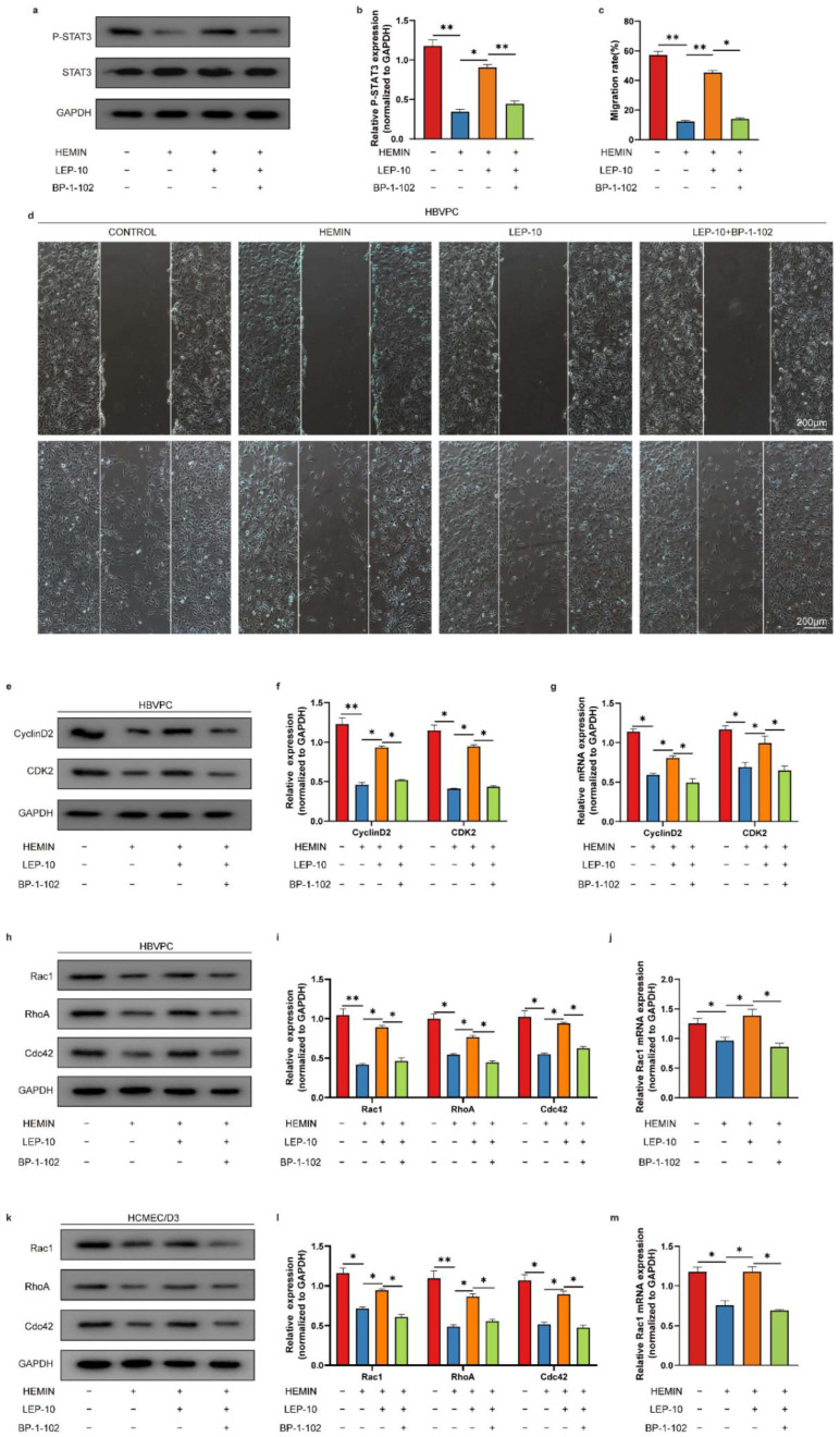
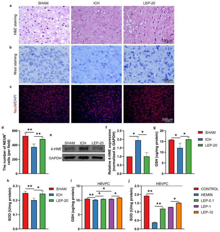
Angiogenesis is a vital endogenous brain self-repair processes for neurological recovery after intracerebral hemorrhage (ICH). Increasing evidence suggests that leptin potentiates angiogenesis and plays a beneficial role in stroke. However, the proangiogenic effect of leptin on ICH has not been adequately explored. Moreover, leptin triggers post-ICH angiogenesis through pericyte, an important component of forming new blood vessels, which remains unclear. Here, we reported that exogenous leptin infusion dose-dependent promoted vascular endothelial cells survival and proliferation at chronic stage of ICH mice. Additionally, leptin robustly ameliorated pericytes loss, enhanced pericytes proliferation and migration in ICH mice in vivo, and in ICH human brain microvascular pericytes (HBVPC) in vitro. Notably, we showed that pericytes-derived pro-angiogenic factors were responsible for enhancing the survival, proliferation and tube formation followed leptin treatment in human brain microvascular endothelial cells (HCMEC/D3)/HBVPC co-culture models. Importantly, considerable improvements in neurobehavioral function and hostile microenvironment were observed in leptin treatment ICH mice, indicating that better vascular functionality post ICH improves outcome. Mechanistically, this study unveiled that leptin boost post-ICH angiogenesis potentially through modulation of leptin receptor (leptinR)/Signal Transducer and Activator of Transcription 3 (STAT3) signaling pathway in pericyte. Thus, leptin may be a lucrative option for the treatment of ICH.

摘要

血管生成是脑出血(ICH)后神经恢复的重要内源性脑自我修复过程。越来越多的证据表明,瘦素促进血管生成,并在中风中发挥有益作用。然而,瘦素对 ICH 的促血管生成作用尚未得到充分探索。此外,瘦素通过周细胞触发 ICH 后的血管生成,周细胞是形成新血管的重要组成部分,其作用机制尚不清楚。在这里,我们报道了外源性瘦素输注在 ICH 小鼠慢性期呈剂量依赖性促进血管内皮细胞的存活和增殖。此外,瘦素在体内显著改善了 ICH 小鼠的周细胞丢失,增强了其增殖和迁移,在体外也增强了 ICH 人脑微血管周细胞(HBVPC)的增殖和迁移。值得注意的是,我们表明周细胞衍生的促血管生成因子是瘦素处理后在人脑血管内皮细胞(HCMEC/D3)/HBVPC 共培养模型中增强细胞存活、增殖和管状形成的原因。重要的是,在接受瘦素治疗的 ICH 小鼠中观察到神经行为功能和恶劣微环境得到了相当大的改善,这表明 ICH 后更好的血管功能可改善预后。从机制上讲,这项研究揭示了瘦素通过调节周细胞中的瘦素受体(leptinR)/信号转导和转录激活因子 3(STAT3)信号通路来促进 ICH 后的血管生成。因此,瘦素可能是治疗 ICH 的一种有前途的选择。

https://cdn.ncbi.nlm.nih.gov/pmc/blobs/55fb/9454866/3fcf9b4bd502/cells-11-02755-g014.jpg
https://cdn.ncbi.nlm.nih.gov/pmc/blobs/55fb/9454866/68053ef3d283/cells-11-02755-g001.jpg
https://cdn.ncbi.nlm.nih.gov/pmc/blobs/55fb/9454866/b017adbd3493/cells-11-02755-g002.jpg
https://cdn.ncbi.nlm.nih.gov/pmc/blobs/55fb/9454866/c48d448bce14/cells-11-02755-g003.jpg
https://cdn.ncbi.nlm.nih.gov/pmc/blobs/55fb/9454866/0fde4ede7c46/cells-11-02755-g004.jpg
https://cdn.ncbi.nlm.nih.gov/pmc/blobs/55fb/9454866/44ed2fbec502/cells-11-02755-g005.jpg
https://cdn.ncbi.nlm.nih.gov/pmc/blobs/55fb/9454866/832a8d46341e/cells-11-02755-g006.jpg
https://cdn.ncbi.nlm.nih.gov/pmc/blobs/55fb/9454866/4feffd7b9bd1/cells-11-02755-g007.jpg
https://cdn.ncbi.nlm.nih.gov/pmc/blobs/55fb/9454866/a69c0bc08fce/cells-11-02755-g008.jpg
https://cdn.ncbi.nlm.nih.gov/pmc/blobs/55fb/9454866/545893bb8f8a/cells-11-02755-g009.jpg
https://cdn.ncbi.nlm.nih.gov/pmc/blobs/55fb/9454866/1753889200e9/cells-11-02755-g010.jpg
https://cdn.ncbi.nlm.nih.gov/pmc/blobs/55fb/9454866/492f5ee3bca0/cells-11-02755-g011.jpg
https://cdn.ncbi.nlm.nih.gov/pmc/blobs/55fb/9454866/5d75b749f7bd/cells-11-02755-g012.jpg
https://cdn.ncbi.nlm.nih.gov/pmc/blobs/55fb/9454866/e4fb42fad4c0/cells-11-02755-g013.jpg
https://cdn.ncbi.nlm.nih.gov/pmc/blobs/55fb/9454866/3fcf9b4bd502/cells-11-02755-g014.jpg

相似文献

1
Leptin Promotes Angiogenesis via Pericyte STAT3 Pathway upon Intracerebral Hemorrhage.瘦素通过脑内出血时的周细胞 STAT3 通路促进血管生成。
Cells. 2022 Sep 3;11(17):2755. doi: 10.3390/cells11172755.
2
Buyang huanwu decoction promotes angiogenesis via vascular endothelial growth factor receptor-2 activation through the PI3K/Akt pathway in a mouse model of intracerebral hemorrhage.补阳还五汤通过PI3K/Akt途径激活血管内皮生长因子受体-2促进脑出血小鼠模型中的血管生成。
BMC Complement Altern Med. 2015 Mar 28;15:91. doi: 10.1186/s12906-015-0605-8.
3
Thrombin promotes pericyte coverage by Tie2 activation in a rat model of intracerebral hemorrhage.凝血酶通过激活大鼠脑出血模型中的 Tie2 促进周细胞覆盖。
Brain Res. 2019 Apr 1;1708:58-68. doi: 10.1016/j.brainres.2018.12.003. Epub 2018 Dec 5.
4
Endothelial PDGF-D contributes to neurovascular protection after ischemic stroke by rescuing pericyte functions.内皮细胞 PDGF-D 通过挽救周细胞功能对缺血性脑卒中后的神经血管保护起作用。
Cell Mol Life Sci. 2024 May 21;81(1):225. doi: 10.1007/s00018-024-05244-w.
5
Leptin combined with withaferin A boost posthemorrhagic neurogenesis via activation of STAT3/SOCS3 pathway.瘦素与醉马草乙素通过激活 STAT3/SOCS3 通路促进脑出血后神经发生。
Exp Neurol. 2024 Jul;377:114809. doi: 10.1016/j.expneurol.2024.114809. Epub 2024 May 5.
6
Excess vascular endothelial growth factor-A disrupts pericyte recruitment during blood vessel formation.过量的血管内皮生长因子 A 会破坏血管生成过程中的周细胞募集。
Angiogenesis. 2019 Feb;22(1):167-183. doi: 10.1007/s10456-018-9648-z. Epub 2018 Sep 20.
7
The adipokine leptin modulates adventitial pericyte functions by autocrine and paracrine signalling.脂联素通过自分泌和旁分泌信号调节外膜周细胞的功能。
Sci Rep. 2017 Jul 14;7(1):5443. doi: 10.1038/s41598-017-05868-y.
8
Hyperlipidemia attenuates vascular endothelial growth factor-induced angiogenesis, impairs cerebral blood flow, and disturbs stroke recovery via decreased pericyte coverage of brain endothelial cells.高脂血症通过减少脑内皮细胞周细胞对血管内皮生长因子诱导的血管生成的覆盖,损害脑血流,并干扰中风恢复。
Arterioscler Thromb Vasc Biol. 2013 Jul;33(7):1561-7. doi: 10.1161/ATVBAHA.112.300749. Epub 2013 Apr 4.
9
Lactate potentiates angiogenesis and neurogenesis in experimental intracerebral hemorrhage.乳酸促进实验性脑出血中的血管生成和神经发生。
Exp Mol Med. 2018 Jul 6;50(7):1-12. doi: 10.1038/s12276-018-0113-2.
10
Leukemia Inhibitory Factor Decreases Neurogenesis and Angiogenesis in a Rat Model of Intracerebral Hemorrhage.白血病抑制因子减少脑出血大鼠模型中的神经发生和血管生成。
Curr Med Sci. 2019 Apr;39(2):298-304. doi: 10.1007/s11596-019-2034-2. Epub 2019 Apr 23.

引用本文的文献

1
Blood-Brain Barrier (BBB) Dysfunction in CNS Diseases: Paying Attention to Pericytes.中枢神经系统疾病中的血脑屏障功能障碍:关注周细胞
CNS Neurosci Ther. 2025 May;31(5):e70422. doi: 10.1111/cns.70422.
2
Rutin ameliorates stress-induced blood‒brain barrier dysfunction and cognitive decline via the endothelial HDAC1‒Claudin-5 axis.芦丁通过内皮组蛋白去乙酰化酶1-闭合蛋白-5轴改善应激诱导的血脑屏障功能障碍和认知能力下降。
Fluids Barriers CNS. 2025 Apr 2;22(1):35. doi: 10.1186/s12987-025-00639-8.
3
Association Between the Expression of T Helper Type 17 Cell-Related Cytokines and Valve Damage in Rheumatic Heart Disease.

本文引用的文献

1
Cardiac pericyte reprogramming by MEK inhibition promotes arteriologenesis and angiogenesis of the ischemic heart.MEK 抑制诱导心脏周细胞重编程促进缺血性心脏的血管生成和动脉生成。
J Clin Invest. 2022 May 16;132(10). doi: 10.1172/JCI152308.
2
Treatment effect of DNA framework nucleic acids on diffuse microvascular endothelial cell injury after subarachnoid hemorrhage.DNA 框架核酸对蛛网膜下腔出血后弥漫性微血管内皮细胞损伤的治疗作用。
Cell Prolif. 2022 Apr;55(4):e13206. doi: 10.1111/cpr.13206. Epub 2022 Feb 21.
3
Hemorrhage-Induced Sphingosine Kinase 1 Contributes to Ferroptosis-Mediated Secondary Brain Injury in Intracerebral Hemorrhage.
17型辅助性T细胞相关细胞因子表达与风湿性心脏病瓣膜损害的关系
Mol Biotechnol. 2024 Nov 22. doi: 10.1007/s12033-024-01321-4.
4
The interactions between energy homeostasis and neurovascular plasticity.能量平衡与神经血管可塑性的相互作用。
Nat Rev Endocrinol. 2024 Dec;20(12):749-759. doi: 10.1038/s41574-024-01021-8. Epub 2024 Jul 25.
5
Mechanisms of Postischemic Stroke Angiogenesis: A Multifaceted Approach.缺血性脑卒中后血管生成的机制:一种多方面的研究方法。
J Inflamm Res. 2024 Jul 12;17:4625-4646. doi: 10.2147/JIR.S461427. eCollection 2024.
6
Immune-mediated disruption of the blood-brain barrier after intracerebral hemorrhage: Insights and potential therapeutic targets.脑出血后血脑屏障的免疫介导破坏:见解与潜在治疗靶点。
CNS Neurosci Ther. 2024 Jul;30(7):e14853. doi: 10.1111/cns.14853.
7
MicroRNA-451 Regulates Angiogenesis in Intracerebral Hemorrhage by Targeting Macrophage Migration Inhibitory Factor.微小 RNA-451 通过靶向巨噬细胞移动抑制因子调节脑出血中的血管生成。
Mol Neurobiol. 2024 Dec;61(12):10481-10499. doi: 10.1007/s12035-024-04207-3. Epub 2024 May 14.
8
Pathological mechanisms and future therapeutic directions of thrombin in intracerebral hemorrhage: a systematic review.凝血酶在脑出血中的病理机制及未来治疗方向:一项系统综述
Front Pharmacol. 2024 Apr 18;15:1293428. doi: 10.3389/fphar.2024.1293428. eCollection 2024.
9
The Role of Cdc42 in the Insulin and Leptin Pathways Contributing to the Development of Age-Related Obesity.Cdc42 在胰岛素和瘦素通路中的作用及其与年龄相关性肥胖发生的关系。
Nutrients. 2023 Nov 29;15(23):4964. doi: 10.3390/nu15234964.
10
EAAT3 impedes oligodendrocyte remyelination in chronic cerebral hypoperfusion-induced white matter injury.兴奋性氨基酸转运体3(EAAT3)在慢性脑灌注不足诱导的白质损伤中阻碍少突胶质细胞的髓鞘再生。
CNS Neurosci Ther. 2024 Jan;30(1):e14487. doi: 10.1111/cns.14487. Epub 2023 Oct 6.
出血诱导的鞘氨醇激酶 1 促进脑出血中铁死亡介导的继发性脑损伤。
Mol Neurobiol. 2022 Mar;59(3):1381-1397. doi: 10.1007/s12035-021-02605-5. Epub 2022 Jan 7.
4
Twenty-Year Change in Severity and Outcome of Ischemic and Hemorrhagic Strokes.二十年来缺血性和出血性中风严重程度和结局的变化。
JAMA Neurol. 2022 Jan 1;79(1):61-69. doi: 10.1001/jamaneurol.2021.4346.
5
Antimicrobial immunity impedes CNS vascular repair following brain injury.抗菌免疫会阻碍脑损伤后的中枢神经系统血管修复。
Nat Immunol. 2021 Oct;22(10):1280-1293. doi: 10.1038/s41590-021-01012-1. Epub 2021 Sep 23.
6
Lessons to be learned in intracerebral haemorrhage research.脑出血研究中可吸取的经验教训。
Lancet Neurol. 2021 Oct;20(10):779-780. doi: 10.1016/S1474-4422(21)00296-9. Epub 2021 Sep 3.
7
Global, regional, and national burden of stroke and its risk factors, 1990-2019: a systematic analysis for the Global Burden of Disease Study 2019.1990—2019年全球、区域和国家的卒中负担及其风险因素:全球疾病负担研究2019的系统分析
Lancet Neurol. 2021 Oct;20(10):795-820. doi: 10.1016/S1474-4422(21)00252-0. Epub 2021 Sep 3.
8
Pericytic porters: helping leptin step in.周细胞搬运工:助瘦素一臂之力。
Trends Endocrinol Metab. 2021 Dec;32(12):958-959. doi: 10.1016/j.tem.2021.08.001. Epub 2021 Aug 19.
9
Leptin receptor-expressing pericytes mediate access of hypothalamic feeding centers to circulating leptin.瘦素受体表达的周细胞介导下丘脑摄食中枢与循环瘦素的接触。
Cell Metab. 2021 Jul 6;33(7):1433-1448.e5. doi: 10.1016/j.cmet.2021.05.017. Epub 2021 Jun 14.
10
Regulation of hippocampal synaptic function by the metabolic hormone leptin: Implications for health and disease.代谢激素瘦素对海马突触功能的调节:对健康和疾病的影响。
Prog Lipid Res. 2021 Apr;82:101098. doi: 10.1016/j.plipres.2021.101098. Epub 2021 Apr 22.