Carrard Johann, Bacher Sebastien, Rochat-Guignard Isabelle, Knebel Jean-François, Alamo Leonor, Meuwly Jean-Yves, Tenisch Estelle
Department of Radiology, Riviera-Chablais Hospital, Rennaz, and University of Lausanne, Lausanne, Switzerland.
Department of Radiology and Interventional Radiology, Lausanne University Hospital and University of Lausanne, Lausanne, Switzerland.
Front Pediatr. 2022 Aug 26;10:898402. doi: 10.3389/fped.2022.898402. eCollection 2022.
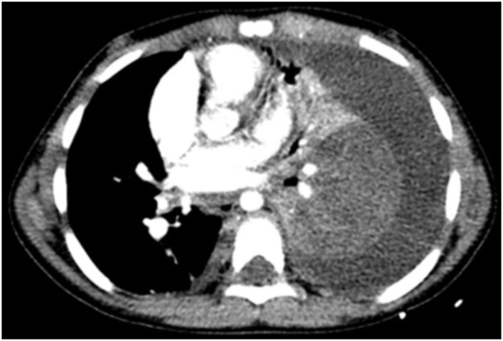
The utilization of contrast-enhanced computed tomography (CT) of the chest for the diagnosis of necrotizing pneumonia (NP), a complication of community-acquired pneumonia, is controversial because of the inherent ionizing radiation involved. Over the past few years, the growing availability of bedside Lung Ultrasound (LUS) devices has led to increased use of this nonionizing imaging method for diagnosing thoracic pathology, including pneumonia.
The objectives of this study were as follows: first, to compare the performance of LUS vs. CT in the identification of certain radiological signs of NP, and second, to determine whether LUS could replace CT in the diagnosis of NP.
We compared retrospectively the CT and LUS images of 41 patients between 2005 and 2018 in whom at least one contrast-injected chest CT scan and one LUS had been undertaken fewer than 7 days apart.
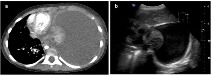
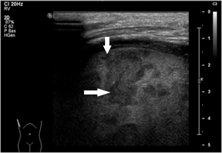
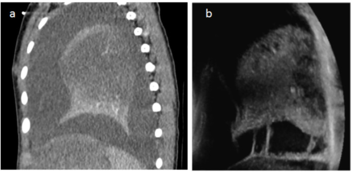
Pleural effusions were demonstrated almost systematically (100% on CT vs. 95.8% on LUS). Visualization of septations in pleural effusions was clearly superior on LUS (20.4% on CT vs 62.5% on LUS). Concerning the detection of necrosis, we observed a strong correlation between LUS and the gold-standard CT (95.8% on LUS vs. 93.7% on CT). Parenchymal cavities were more easily detected on CT than on LUS (79.1 vs. 35.4%).
LUS has shown to be as effective as CT in the diagnosis of NP. The use of CT in patients with NP could be limited to the detection of complications such as bronchopleural fistulae in unfavorably evolving diseases.
胸部对比增强计算机断层扫描(CT)用于诊断社区获得性肺炎的并发症坏死性肺炎(NP)存在争议,因为其涉及电离辐射。在过去几年中,床边肺部超声(LUS)设备的日益普及导致这种非电离成像方法在诊断包括肺炎在内的胸部病变中的使用增加。
本研究的目的如下:第一,比较LUS与CT在识别NP某些放射学征象方面的性能;第二,确定LUS在NP诊断中是否可以替代CT。
我们回顾性比较了2005年至2018年间41例患者的CT和LUS图像,这些患者在间隔不到7天的时间内至少进行了一次胸部增强CT扫描和一次LUS检查。
几乎所有患者均显示有胸腔积液(CT显示率为100%,LUS显示率为95.8%)。LUS对胸腔积液中分隔的显示明显优于CT(CT显示率为20.4%,LUS显示率为62.5%)。关于坏死的检测,我们观察到LUS与金标准CT之间有很强的相关性(LUS显示率为95.8%,CT显示率为93.7%)。CT比LUS更容易检测到实质空洞(分别为79.1%和35.4%)。
LUS在NP诊断中已显示出与CT同样有效。NP患者使用CT可能仅限于检测病情进展不利时的并发症,如支气管胸膜瘘。