Li Ze-Yang, Ma Teng, Yu Ying, Hu Bo, Han Yu, Xie Hao, Ni Min-Hua, Chen Zhu-Hong, Zhang Yang-Ming, Huang Yu-Xiang, Li Wen-Hua, Wang Wen, Yan Lin-Feng, Cui Guang-Bin
Department of Radiology, Functional and Molecular Imaging Key Lab of Shaanxi Province, Tangdu Hospital, Fourth Military Medical University, Xi'an, China.
Faculty of Medical Technology, Shaanxi University of Chinese Medicine, Xianyang, China.
Front Neurol. 2022 Aug 24;13:923310. doi: 10.3389/fneur.2022.923310. eCollection 2022.
Neuroimaging meta-analysis identified abnormal neural activity alterations in patients with type 2 diabetes mellitus (T2DM), but there was no consistency or heterogeneity analysis between different brain imaging processing strategies. The aim of this meta-analysis was to determine consistent changes of regional brain functions in T2DM the indicators obtained by using different post-processing methods.
Since the indicators obtained using varied post-processing methods reflect different neurophysiological and pathological characteristics, we further conducted a coordinate-based meta-analysis (CBMA) of the two categories of neuroimaging literature, which were grouped according to similar data processing methods: one group included regional homogeneity (ReHo), independent component analysis (ICA), and degree centrality (DC) studies, while the other group summarized the literature on amplitude of low-frequency fluctuation (ALFF) and cerebral blood flow (CBF).
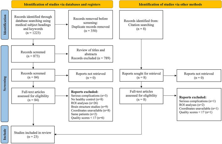
The final meta-analysis included 23 eligible trials with 27 data sets. Compared with the healthy control group, when neuroimaging studies were combined with ReHo, ICA, and DC measurements, the brain activity of the right Rolandic operculum, right supramarginal gyrus, and right superior temporal gyrus in T2DM patients decreased significantly. When neuroimaging studies were combined with ALFF and CBF measurements, there was no clear evidence of differences in the brain function between T2DM and HCs.
T2DM patients have a series of spontaneous abnormal brain activities, mainly involving brain regions related to learning, memory, and emotion, which provide early biomarkers for clarifying the mechanism of cognitive impairment and neuropsychiatric disorders in diabetes.
https://www.crd.york.ac.uk/prospero/display_record.php?RecordID=247071, PROSPERO [CRD42021247071].
神经影像学荟萃分析发现2型糖尿病(T2DM)患者存在异常神经活动改变,但不同脑成像处理策略之间未进行一致性或异质性分析。本荟萃分析的目的是确定T2DM患者使用不同后处理方法获得的指标下脑区功能的一致性变化。
由于使用不同后处理方法获得的指标反映了不同的神经生理和病理特征,我们进一步对两类神经影像学文献进行了基于坐标的荟萃分析(CBMA),这两类文献根据相似的数据处理方法进行分组:一组包括局部一致性(ReHo)、独立成分分析(ICA)和度中心性(DC)研究,另一组汇总了低频振幅(ALFF)和脑血流量(CBF)的文献。
最终的荟萃分析纳入了23项符合条件的试验,共27个数据集。与健康对照组相比,当神经影像学研究结合ReHo、ICA和DC测量时,T2DM患者右侧中央前回、右侧缘上回和右侧颞上回的脑活动显著降低。当神经影像学研究结合ALFF和CBF测量时,没有明确证据表明T2DM患者与健康对照者之间脑功能存在差异。
T2DM患者存在一系列自发性异常脑活动,主要涉及与学习、记忆和情感相关的脑区,这为阐明糖尿病认知障碍和神经精神疾病的机制提供了早期生物标志物。
https://www.crd.york.ac.uk/prospero/display_record.php?RecordID=247071,PROSPERO [CRD42021247071] 。