Liu Jieke, Li Yong, Yang Xi, Xu Hao, Ren Jing, Zhou Peng
Department of Radiology, Sichuan Cancer Hospital and Institute, Sichuan Cancer Center, School of Medicine, University of Electronic Science and Technology of China, Chengdu, China.
Front Aging Neurosci. 2021 Jun 17;13:678359. doi: 10.3389/fnagi.2021.678359. eCollection 2021.
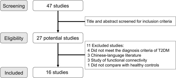
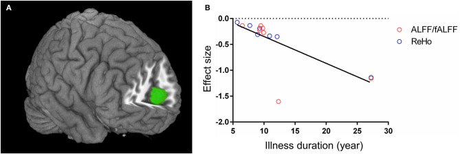
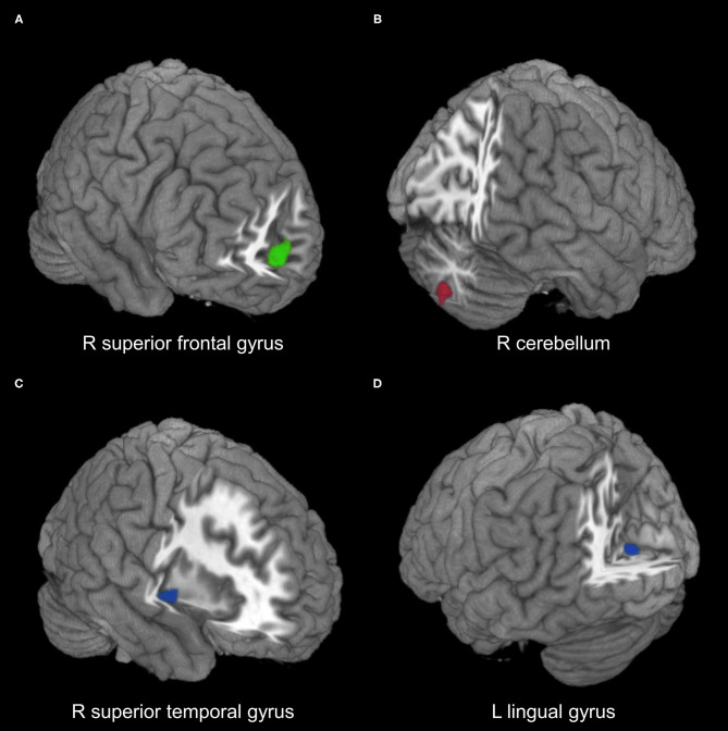
Resting-state functional magnetic resonance imaging (rs-fMRI) studies have revealed inconsistent regional spontaneous neural activity alterations in patients with type 2 diabetes mellitus (T2DM). The aim of our meta-analysis was to identify concordant regional spontaneous neural activity abnormalities in patients with T2DM. A systematic search was conducted to identify voxel-based rs-fMRI studies comparing T2DM patients with healthy controls. The permutation of subject images seed-based mapping (SDM) was used to quantitatively estimate the regional spontaneous neural activity abnormalities in patients with T2DM. Metaregression was conducted to examine the associations between clinical characteristics and functional alterations. A total of 16 studies with 19 datasets including 434 patients with T2DM and 391 healthy controls were included. Patients with T2DM showed hypoactivity in the right medial superior frontal gyrus, right superior temporal gyrus, and left lingual gyrus, whereas hyperactivity in the right cerebellum. Metaregression analysis identified negative correlation between regional activity in the medial superior frontal and anterior cingulate gyri and illness duration of patients with T2DM. The patterns of regional spontaneous neural activity alterations, characterized by hypoactivity in the medial pre-frontal cortex, visual cortex, and superior temporal gyrus, whereas hyperactivity in the cerebellum, might represent the underlying neuropathological mechanisms of T2DM.
静息态功能磁共振成像(rs-fMRI)研究显示,2型糖尿病(T2DM)患者区域自发性神经活动改变存在不一致性。我们进行荟萃分析的目的是确定T2DM患者一致的区域自发性神经活动异常。我们进行了系统检索,以识别基于体素的rs-fMRI研究,这些研究比较了T2DM患者与健康对照。采用基于种子点映射的受试者图像置换(SDM)来定量评估T2DM患者区域自发性神经活动异常。进行元回归分析以检验临床特征与功能改变之间的关联。共纳入16项研究的19个数据集,包括434例T2DM患者和391例健康对照。T2DM患者右侧额上回中部、右侧颞上回和左侧舌回表现为活动减退,而右侧小脑表现为活动增强。元回归分析确定,额上回中部和前扣带回区域活动与T2DM患者病程呈负相关。以内侧前额叶皮质、视觉皮质和颞上回活动减退,而小脑活动增强为特征的区域自发性神经活动改变模式,可能代表了T2DM潜在的神经病理机制。