Badawy Mohamed, Johnson Geoffrey, Jain Manoj, Kendi Ayse Tuba, Johnson Derek R, Porter Alyx, Yang Ming, Sonbol Mohamad Bassam
Division of Radiology and Nuclear Medicine, Mayo Clinic, Rochester, MN.
Division of Radiology and Nuclear Medicine, Mayo Clinic, Jacksonville, Florida, USA.
BJR Case Rep. 2022 Jan 27;8(3):20210222. doi: 10.1259/bjrcr.20210222. eCollection 2022 Sep.
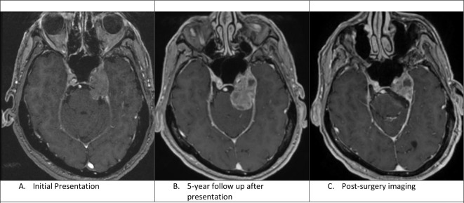
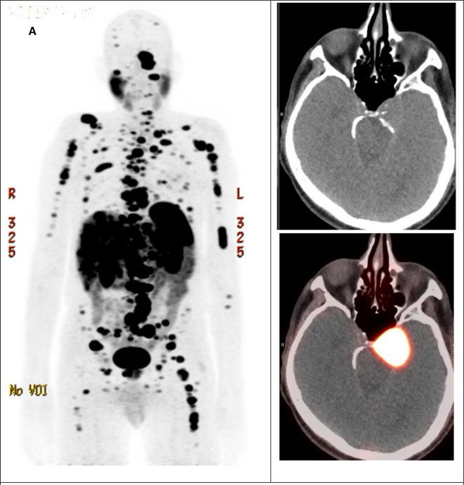
Metastatic neuroendocrine tumour (NET) to brain has been reported in 1.5-5% of patients with NETs. Differentiation between intracranial NET metastasis and meningiomas can cause a diagnostic dilemma. We present a symptomatic case of a 66-year-old male with a history of left-sided skull base mass. The diagnosis of a meningioma was made based on the MRI findings and clinical presentation. The patient received radiation and the mass remained stable on serial MRI images at follow-up visits. Five years after his initial presentation, the patient's mass showed further growth. He also complained of worsening of his recent diagnosis of irritable bowel syndrome and fluctuations in his blood pressure. Surgical resection was performed, and histopathological features were consistent with moderately differentiated neuroendocrine tumour. Further evaluation with 68 Gallium-DOTATATE positron emission-computed tomography (Ga-68 PET/CT) showed metastatic disease involving the bones, lymph nodes, and liver without convincing evidence of the location of primary malignancy within the bowel loops or the pancreas. The patient was started on combination of capecitabine and temozolomide with partial response and significant improvement of his symptoms. This case highlights the clinical and radiological behaviour of intracranial NET that can mimic the diagnosis of meningioma.
据报道,1.5%-5%的神经内分泌肿瘤(NET)患者会发生脑转移。颅内NET转移瘤与脑膜瘤的鉴别诊断可能会造成诊断难题。我们报告一例有症状的66岁男性病例,其有左侧颅底肿物病史。根据磁共振成像(MRI)检查结果及临床表现诊断为脑膜瘤。患者接受了放疗,随访期间系列MRI图像显示肿物稳定。初次就诊5年后,患者的肿物进一步增大。他还主诉近期诊断的肠易激综合征病情加重以及血压波动。进行了手术切除,组织病理学特征符合中度分化神经内分泌肿瘤。进一步行68镓-奥曲肽正电子发射断层扫描-计算机断层扫描(Ga-68 PET/CT)检查显示,骨、淋巴结和肝脏有转移病灶,但未发现肠袢或胰腺内有原发性恶性肿瘤的确切证据。患者开始接受卡培他滨和替莫唑胺联合治疗,部分缓解,症状明显改善。该病例突出了颅内NET可模仿脑膜瘤诊断的临床和影像学表现。