Ivonin Alexey G, Smirnova Svetlana L, Roshchevskaya Irina M
Department of Comparative Cardiology - Komi Scientific Centre of the Ural Branch of the Russian Academy of Sciences , Syktyvkar - Federação Russa.
Laboratory of Pharmacological Screening - Research Zakusov Institute of Pharmacology , Moscow - Federação Russa.
Arq Bras Cardiol. 2022 Sep 12;119(5):766-75. doi: 10.36660/abc.20211058.
Exhaustive physical exercise can cause substantial changes in the electrical properties of the myocardium.
To evaluate, using body surface potential mapping, the electrical activity of the heart in rats during ventricular depolarization after acute exhaustive exercise.
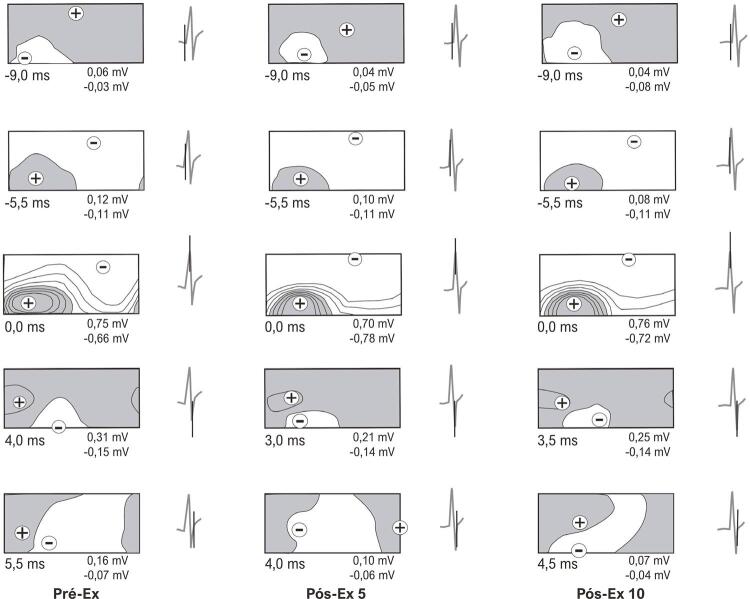
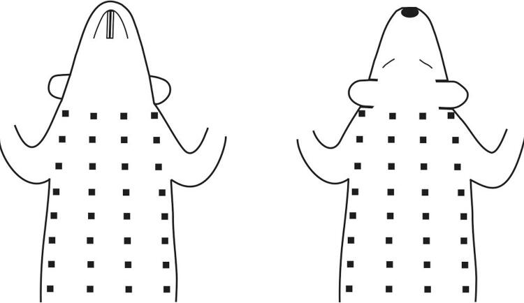
Twelve-week-old male rats were submitted to acute treadmill exercise at 36 m/min until exhaustion. Unipolar electrocardiograms (ECGs) from the torso surface were recorded in zoletil-anesthetized rats three to five days before (Pre-Ex), 5 and 10 minutes after exhaustive exercise (Post-Ex 5 and Post-Ex 10, respectively) simultaneously with ECGs in limb leads. The instantaneous body surface potential maps (BSPMs) were analyzed during ventricular depolarization. P values <0.05 were considered statistically significant.
Compared with Pre-Ex, an early completion of the second inversion of potential distributions, an early completion of ventricular depolarization, as well as a decrease in the duration of the middle phase and the total duration of ventricular depolarization on BSPMs were revealed at Post-Ex 5. Also, compared with Pre-Ex, an increase in the amplitude of negative BSPM extremum at the R-wave peak on the ECG in lead II (RII-peak) and a decrease in the amplitude of negative BSPM extremum at 3 and 4 ms after RII-peak were showed at Post-Ex 5. At Post-Ex 10, parameters of BSPMs did not differ from those at Pre-Ex.
In rats, acute exhaustive exercise causes reversible changes in the temporal and amplitude characteristics of BSPMs during ventricular depolarization, most likely related to alterations in the excitation of the main mass of the ventricular myocardium.
力竭性体育运动会导致心肌电特性发生显著变化。
使用体表电位标测技术评估急性力竭运动后大鼠心室去极化过程中心脏的电活动。
12周龄雄性大鼠以36米/分钟的速度在跑步机上进行急性运动直至力竭。在力竭运动前3至5天(运动前)、力竭运动后5分钟和10分钟(分别为运动后5分钟和运动后10分钟),对用佐替卡因麻醉的大鼠记录其躯干表面的单极心电图(ECG),同时记录肢体导联的ECG。在心室去极化过程中分析瞬时体表电位图(BSPM)。P值<0.05被认为具有统计学意义。
与运动前相比,运动后5分钟时,体表电位分布第二次反转提前完成,心室去极化提前完成,且体表电位图上心室内期持续时间和心室去极化总持续时间缩短。此外,与运动前相比,运动后5分钟时,II导联心电图R波峰(RII峰)处体表电位图负向极值的幅度增加,RII峰后3毫秒和4毫秒处体表电位图负向极值的幅度降低。运动后10分钟时,体表电位图参数与运动前无差异。
在大鼠中,急性力竭运动会导致心室去极化过程中体表电位图的时间和幅度特征发生可逆性变化,最可能与心室心肌主要部分的兴奋改变有关。