Department of Pathology and Laboratory Medicine, David Geffen School of Medicine at UCLA, 10833 Le Conte Avenue, 13-145 CHS, BOX 951732, Los Angeles, CA, 90095-1732, USA.
Department of Biostatistics, Fielding School of Public Health at UCLA, Los Angeles, CA, USA.
Diagn Pathol. 2022 Sep 14;17(1):70. doi: 10.1186/s13000-022-01251-2.
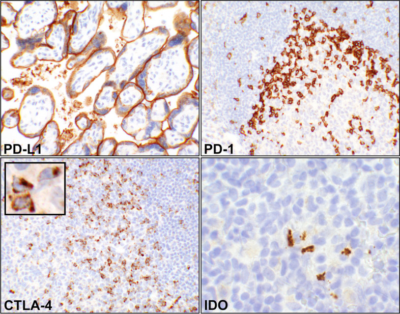
Immune checkpoints including programmed death-ligand 1/programmed death-1/ (PD-L1/PD-1), cytotoxic T lymphocyte antigen 4 (CTLA-4), and indolaimine-2, 3-deoxygenase (IDO) have recently emerged as effective candidates for treatment against a range of human malignancies. We have investigated their expression in the uterine mesenchymal tumors.
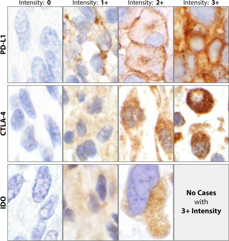
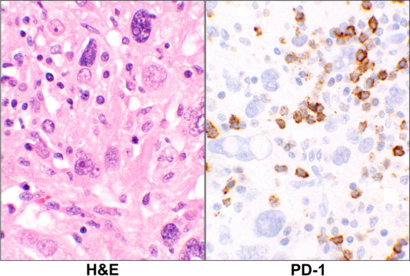
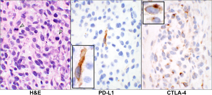
Sixty-eight mesenchymal tumors were categorized into 6 diagnostic groups. We assessed PD-L1, PD-1, CTLA-4, and IDO expression on paraffin embedded tissue blocks of the uterine tumors using the respective antibodies. Immunohistochemical (IHC) stains were classified as positive when the reactions were present in at least 1% of the cell membranes for PD-L1/PD-1 or in cytoplasm for CTLA-4 and IDO, regardless of intensity. Student's t-test and McNemar's chi-square tests were carried out to analyze the results.
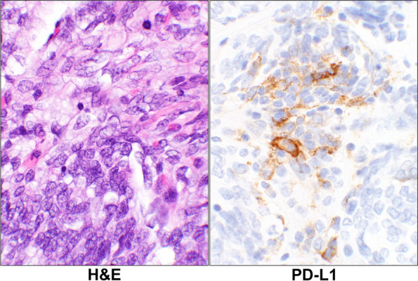
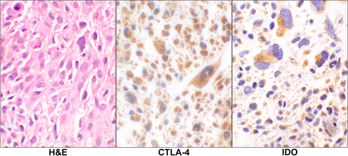
The mesenchymal neoplasms had expressed the immune checkpoints in the tumor and/or the lymphoid cells at the rate of 49% and 54% respectively. The tumor cells were positive in 10 (18%, PD-L1), 0 (0%, PD-1), 18 (32%, CTLA-4), and 13 (23%, IDO) cases while the infiltrating lymphoid cells were positive in 10 (18%, PD-L1), 23 (40%, PD-1), 18 (32%, CTLA-4), and 13 (23%, IDO) cases. Overall, comparison of paired tumor vs lymphoid cells resulted in p-values of ≤ 0.04.
Nearly 50% of the uterine tumors express at least one of the immune checkpoints in tumor and/or the infiltrating lymphoid cells. However, expression of the proteins in the two cellular components are mutually exclusive. Namely, when tumor cells express an immune checkpoint, the infiltrating lymphoid cells do not, and vice versa. Since the leiomyosarcomas are reportedly resistant to the immunotherapy when PD-L1 is expressed in the tumor cells, it can be posited that presence of the IHC positive lymphoid cells may be a better indicator of response to the treatment.
免疫检查点,包括程序性死亡配体 1/程序性死亡-1/(PD-L1/PD-1)、细胞毒性 T 淋巴细胞抗原 4(CTLA-4)和吲哚胺 2,3-双加氧酶(IDO),最近已成为治疗多种人类恶性肿瘤的有效候选药物。我们研究了它们在子宫间质性肿瘤中的表达。
将 68 个间质性肿瘤分为 6 个诊断组。我们使用相应的抗体评估 PD-L1、PD-1、CTLA-4 和 IDO 在子宫肿瘤石蜡包埋组织块上的表达。PD-L1/PD-1 的细胞膜上至少有 1%存在反应,或 CTLA-4 和 IDO 的细胞质中存在反应,无论强度如何,免疫组织化学(IHC)染色均被归类为阳性。进行了学生 t 检验和 McNemar 卡方检验来分析结果。
间质性肿瘤以 49%和 54%的比例分别在肿瘤和/或淋巴细胞中表达免疫检查点。肿瘤细胞阳性 10 例(18%,PD-L1)、0 例(0%,PD-1)、18 例(32%,CTLA-4)和 13 例(23%,IDO),而浸润性淋巴细胞阳性 10 例(18%,PD-L1)、23 例(40%,PD-1)、18 例(32%,CTLA-4)和 13 例(23%,IDO)。总体而言,肿瘤与浸润性淋巴细胞配对比较的 p 值≤0.04。
近 50%的子宫肿瘤在肿瘤和/或浸润性淋巴细胞中表达至少一种免疫检查点。然而,两种细胞成分中的蛋白表达是相互排斥的。即当肿瘤细胞表达免疫检查点时,浸润性淋巴细胞不表达,反之亦然。由于报道称当肿瘤细胞中表达 PD-L1 时,平滑肌肉瘤对免疫治疗有抵抗力,因此可以假设 IHC 阳性淋巴细胞的存在可能是对治疗反应的更好指标。