• 文献检索
  • 文档翻译
  • 深度研究
  • 学术资讯
  • Suppr Zotero 插件Zotero 插件
  • 邀请有礼
  • 套餐&价格
  • 历史记录
应用&插件
Suppr Zotero 插件Zotero 插件浏览器插件Mac 客户端Windows 客户端微信小程序
定价
高级版会员购买积分包购买API积分包
服务
文献检索文档翻译深度研究API 文档MCP 服务
关于我们
关于 Suppr公司介绍联系我们用户协议隐私条款
关注我们

Suppr 超能文献

核心技术专利:CN118964589B侵权必究
粤ICP备2023148730 号-1Suppr @ 2026

文献检索

告别复杂PubMed语法,用中文像聊天一样搜索,搜遍4000万医学文献。AI智能推荐,让科研检索更轻松。

立即免费搜索

文件翻译

保留排版,准确专业,支持PDF/Word/PPT等文件格式,支持 12+语言互译。

免费翻译文档

深度研究

AI帮你快速写综述,25分钟生成高质量综述,智能提取关键信息,辅助科研写作。

立即免费体验

导管导向计算机断层扫描血管造影术:图文综述

Catheter-directed computed tomography angiography: A pictorial essay.

作者信息

Ghosh Abheek, Moxley Ellen, Waghmarae Suneet, Stoner James, Anand Sheena, Akhter Nabeel M

机构信息

Department of Interventional Radiology, University of Maryland School of Medicine, Baltimore, Maryland, United States.

Department of Radiology, University of Maryland, Baltimore, Maryland, United States.

出版信息

J Clin Imaging Sci. 2022 Aug 18;12:49. doi: 10.25259/JCIS_76_2022. eCollection 2022.

DOI:10.25259/JCIS_76_2022
PMID:36128352
原文链接:https://pmc.ncbi.nlm.nih.gov/articles/PMC9479526/
Abstract

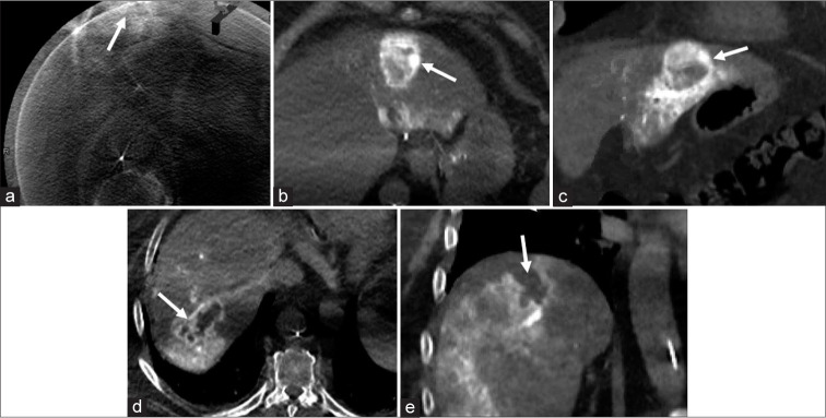
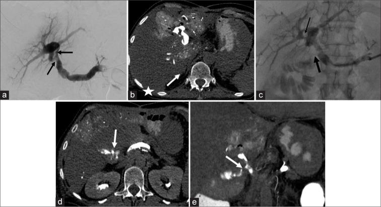
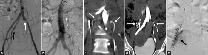
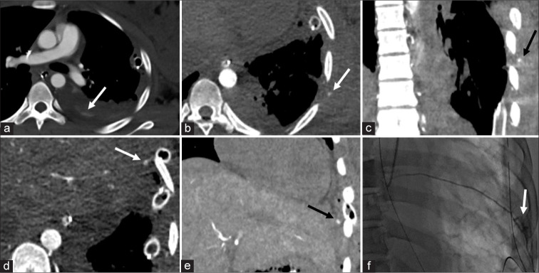
Catheter-directed computed tomography angiography (CDCTA) is an imaging technique where CT images are acquired after selective catheterization of a vessel. Images obtained in this fashion provide several advantages over conventional imaging techniques such as fluoroscopic angiography, digital subtraction angiography, cone-beam CT, and conventional CT angiography. At this point, there is still limited literature on the subject, with prior studies examining a small number of potential uses. The goal of this pictorial essay is to illustrate our single tertiary care center experience using CDCTA.

摘要

导管导向计算机断层血管造影(CDCTA)是一种成像技术,即在对血管进行选择性导管插入术后获取CT图像。以这种方式获得的图像比传统成像技术(如荧光透视血管造影、数字减影血管造影、锥形束CT和传统CT血管造影)具有若干优势。目前,关于该主题的文献仍然有限,先前的研究仅考察了少数潜在用途。本文的目的是阐述我们在单一三级医疗中心使用CDCTA的经验。

https://cdn.ncbi.nlm.nih.gov/pmc/blobs/6d57/9479526/4209e26f3d68/JCIS-12-49-g005.jpg
https://cdn.ncbi.nlm.nih.gov/pmc/blobs/6d57/9479526/234db75c00ca/JCIS-12-49-g001.jpg
https://cdn.ncbi.nlm.nih.gov/pmc/blobs/6d57/9479526/ef4ba8db2c86/JCIS-12-49-g002.jpg
https://cdn.ncbi.nlm.nih.gov/pmc/blobs/6d57/9479526/33720867b8bc/JCIS-12-49-g003.jpg
https://cdn.ncbi.nlm.nih.gov/pmc/blobs/6d57/9479526/bc6b4a86cb91/JCIS-12-49-g004.jpg
https://cdn.ncbi.nlm.nih.gov/pmc/blobs/6d57/9479526/4209e26f3d68/JCIS-12-49-g005.jpg

相似文献

1
Catheter-directed computed tomography angiography: A pictorial essay.导管导向计算机断层扫描血管造影术:图文综述
J Clin Imaging Sci. 2022 Aug 18;12:49. doi: 10.25259/JCIS_76_2022. eCollection 2022.
2
Analysis of enlarged images using time-of-flight magnetic resonance angiography, computed tomography, and conventional angiography.使用飞行时间磁共振血管造影、计算机断层扫描和传统血管造影对放大图像进行分析。
J Med Syst. 2014 Dec;38(12):146. doi: 10.1007/s10916-014-0146-6. Epub 2014 Oct 29.
3
Frameless Co-Registration of Biplane 2D Digital Subtraction Angiography Whole Frames and 3D Rotational Angiography-Based Cone-Beam Computed Tomography Angiogram on Dedicated Software for Stereotactic Radiosurgery of Cranial Vascular Malformations.基于专用软件的双平面二维数字减影血管造影全帧与三维旋转血管造影锥束计算机断层扫描血管造影在颅内血管畸形立体定向放射外科中的无框架配准
Cureus. 2022 Aug 14;14(8):e27983. doi: 10.7759/cureus.27983. eCollection 2022 Aug.
4
Flow Diverter Apposition in Patients with Large or Giant Intracranial Aneurysms Evaluated on Three-Dimensional Fusion Images Acquired by High-Resolution Cone-Beam Computed Tomography and Digital Subtraction Angiography.高分辨率锥形束 CT 和数字减影血管造影三维融合图像评估的大型或巨大颅内动脉瘤患者的血流导向装置贴附。
World Neurosurg. 2021 Mar;147:e388-e395. doi: 10.1016/j.wneu.2020.12.068. Epub 2021 Jan 7.
5
Hybrid angiography-CT for transarterial radioembolization: a pictorial essay.杂交血管造影-CT 在经动脉放射性栓塞治疗中的应用:影像学研究。
Abdom Radiol (NY). 2021 Jun;46(6):2850-2854. doi: 10.1007/s00261-020-02914-8. Epub 2021 Jan 4.
6
Ultra-low-dose intra-arterial contrast injection for iliofemoral computed tomographic angiography.超低位动脉内对比剂注射在髂股动脉 CT 血管造影中的应用。
JACC Cardiovasc Imaging. 2009 Dec;2(12):1404-11. doi: 10.1016/j.jcmg.2009.08.010.
7
C-arm cone-beam CT: general principles and technical considerations for use in interventional radiology.C形臂锥形束CT:介入放射学应用的一般原理和技术考量
J Vasc Interv Radiol. 2009 Jul;20(7 Suppl):S538-44. doi: 10.1016/j.jvir.2009.04.026.
8
[Study of the liver and the portal venous system with digital rotational angiography].[数字旋转血管造影对肝脏和门静脉系统的研究]
Radiol Med. 2001 Mar;101(3):118-24.
9
Usefulness of cone-beam volume CT with flat panel detectors in conjunction with catheter angiography for transcatheter arterial embolization.平板探测器锥形束容积CT联合导管血管造影在经导管动脉栓塞术中的应用价值
J Vasc Interv Radiol. 2007 Dec;18(12):1508-16. doi: 10.1016/j.jvir.2007.08.003.
10
Value of C-Arm Cone Beam Computed Tomography Image Fusion in Maximizing the Versatility of Endovascular Robotics.C型臂锥形束计算机断层扫描图像融合在最大化血管内机器人多功能性方面的价值
Ann Vasc Surg. 2016 Jan;30:138-48. doi: 10.1016/j.avsg.2015.06.070. Epub 2015 Aug 6.

本文引用的文献

1
Catheter-Directed Computed Tomography Hepatic Angiography for Yttrium-90 Selective Internal Radiotherapy of Hepatocellular Carcinoma Reduces Prophylactic Embolization of Extrahepatic Vessels.经导管计算机断层扫描肝动脉造影行钇-90 选择性内放射治疗肝细胞癌可减少肝外血管预防性栓塞。
Cardiovasc Intervent Radiol. 2020 Mar;43(3):478-487. doi: 10.1007/s00270-019-02362-y. Epub 2019 Nov 8.
2
Ultralow Iodine Dose Transarterial Catheter-Directed CT Angiography for Fenestrated Endovascular Aortic Repair Planning.超低碘剂量经动脉导管导向CT血管造影术用于开窗式血管腔内主动脉修复术规划
Ann Vasc Surg. 2016 Aug;35:234-7. doi: 10.1016/j.avsg.2016.01.045. Epub 2016 May 27.
3
Intra-arterial Ultra-low-Dose CT Angiography of Lower Extremity in Diabetic Patients.
糖尿病患者下肢动脉超低剂量CT血管造影
Cardiovasc Intervent Radiol. 2016 Aug;39(8):1165-9. doi: 10.1007/s00270-016-1358-6. Epub 2016 May 5.