Biochemistry Department, School of Medicine and Biomedical Sciences, State University of New York at Buffalo, Buffalo, NY, United States.
Department of Biology, Indiana University, Bloomington, IN, United States.
Front Endocrinol (Lausanne). 2022 Sep 5;13:932286. doi: 10.3389/fendo.2022.932286. eCollection 2022.
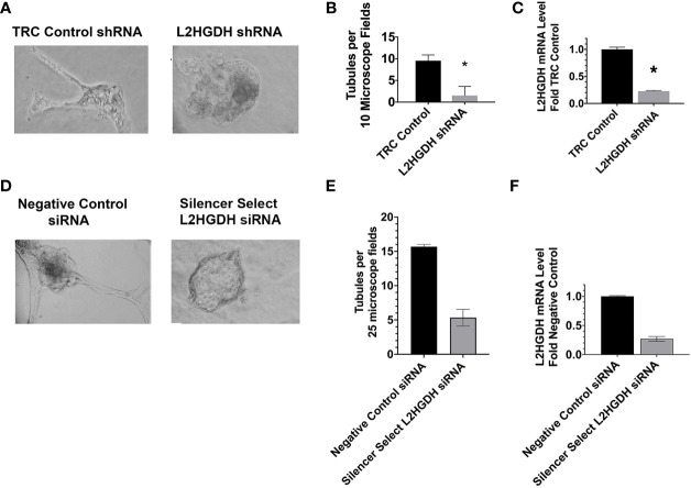
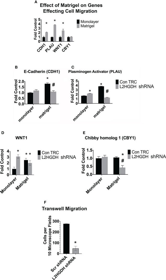
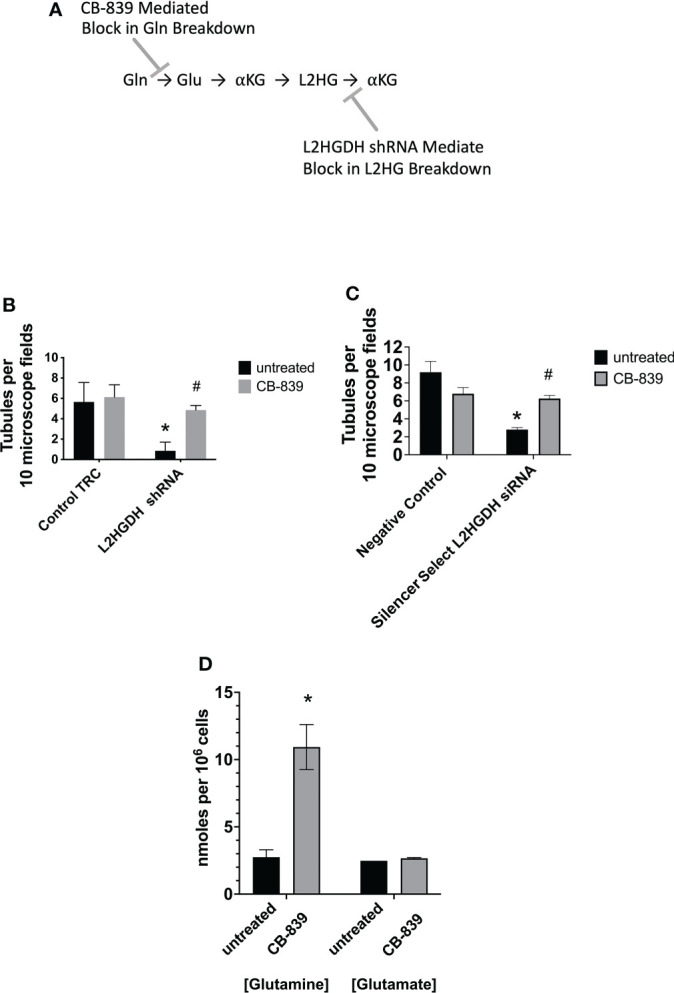
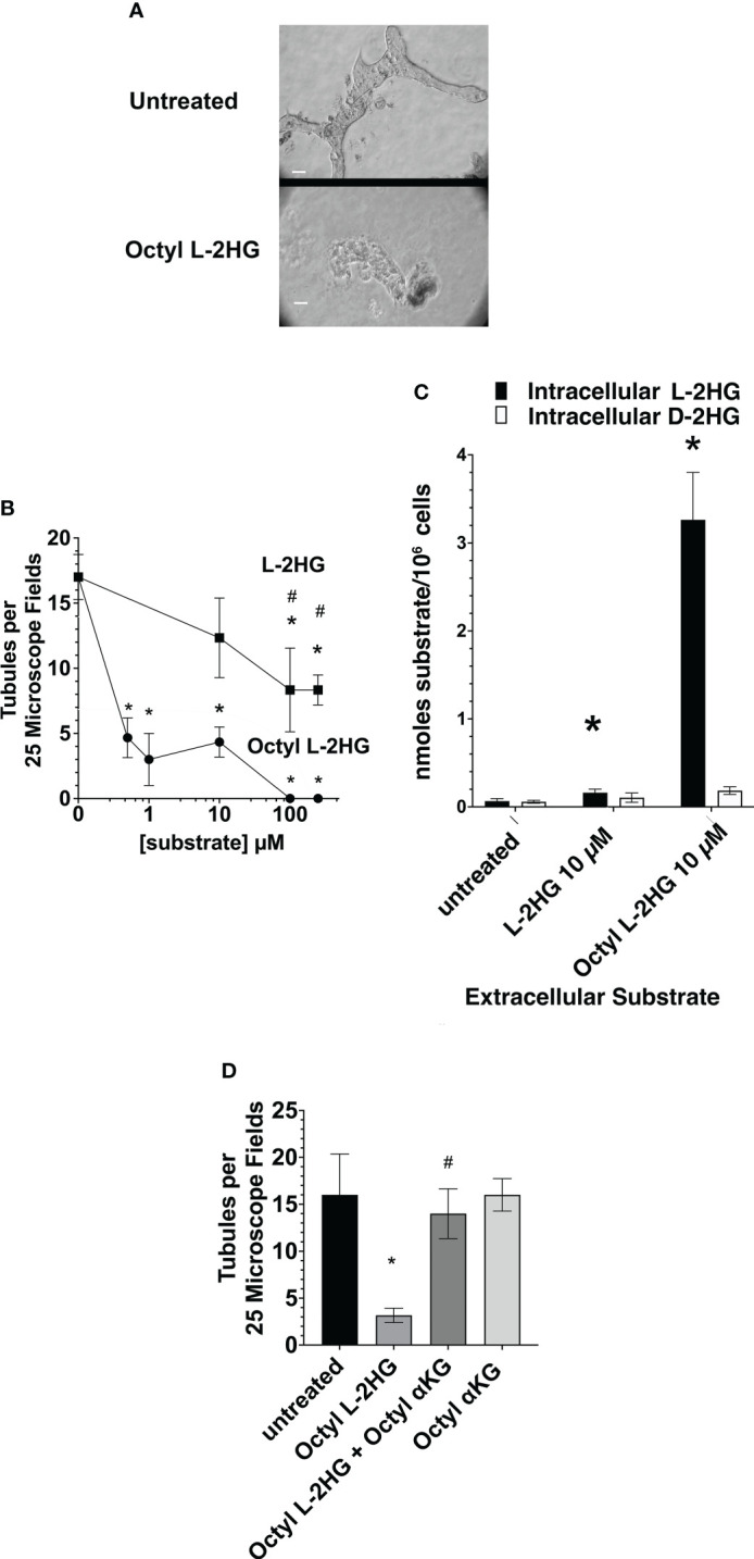
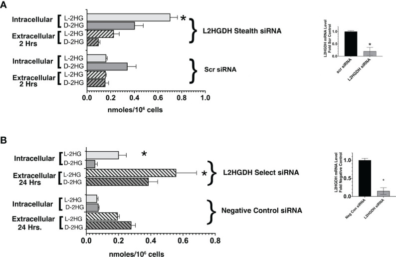
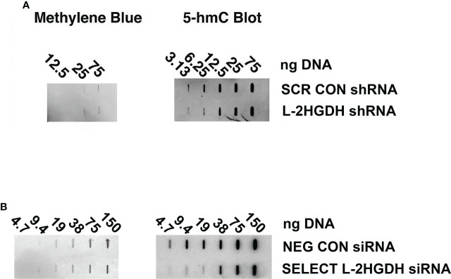
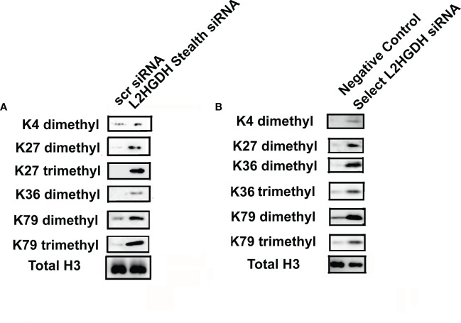
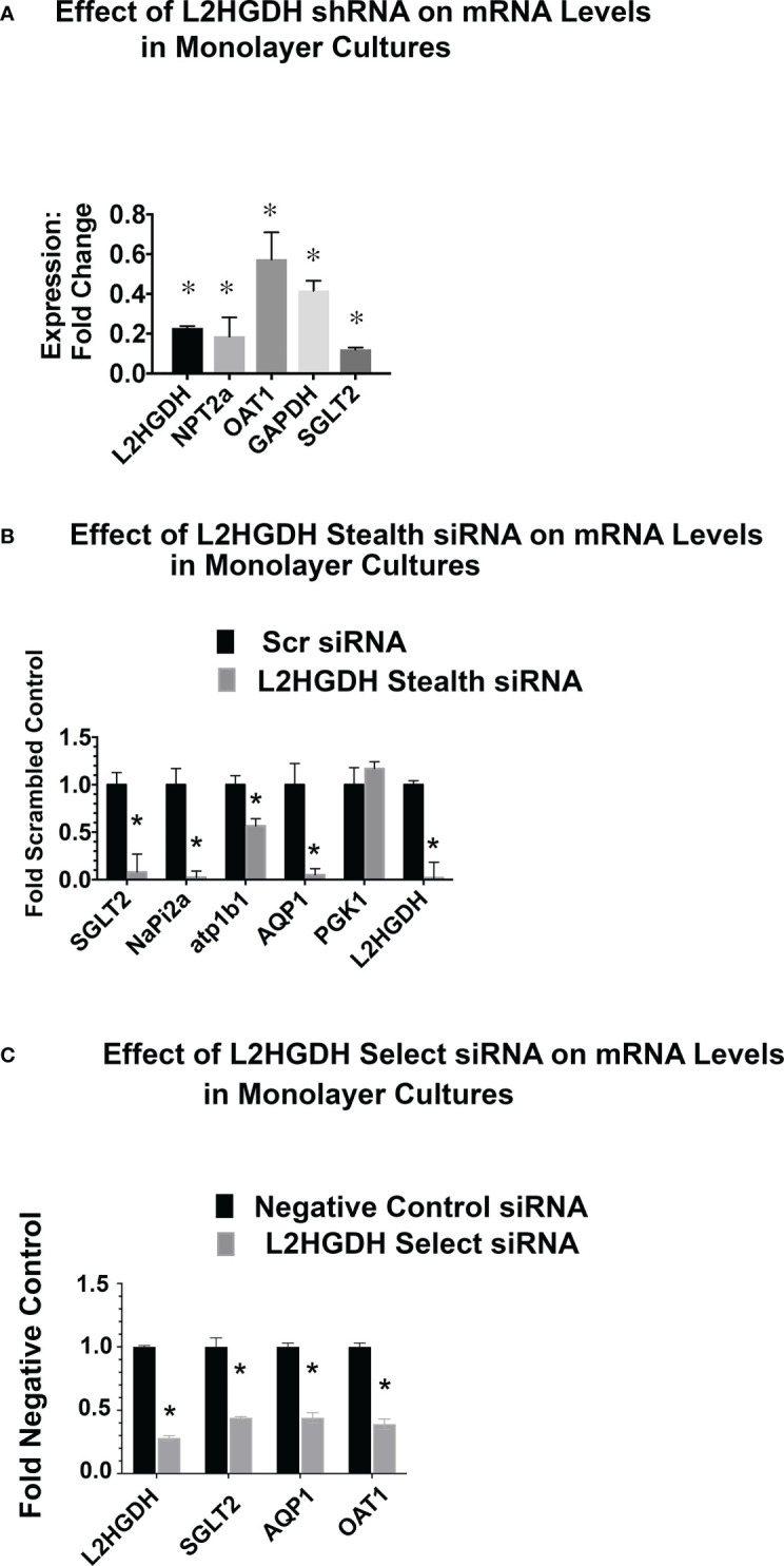
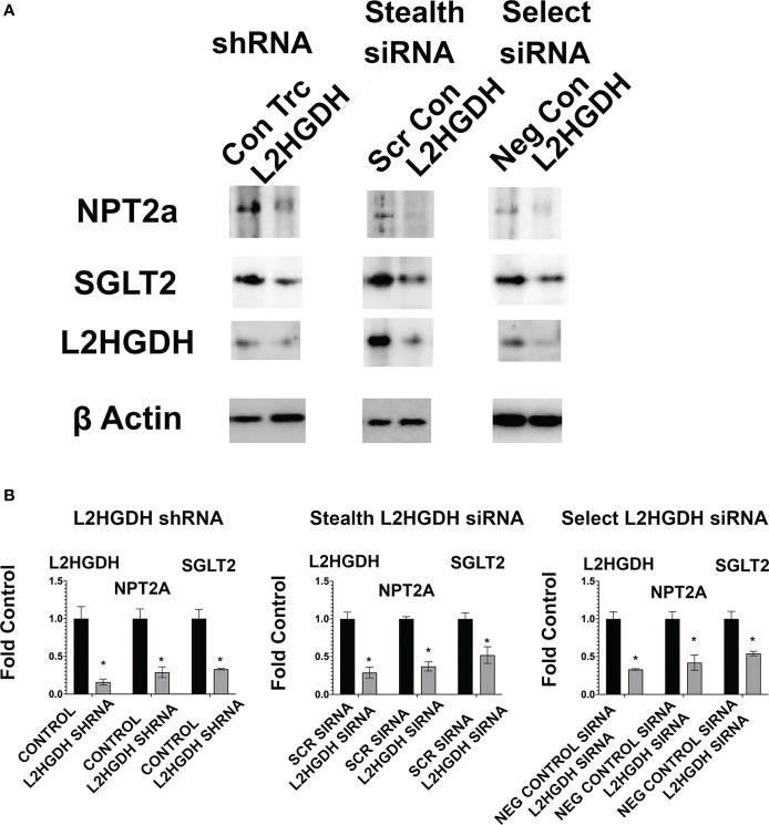
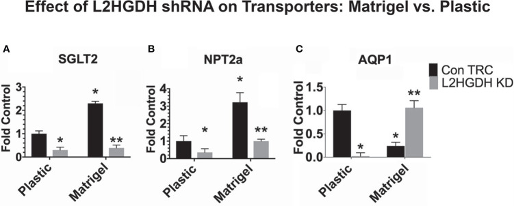
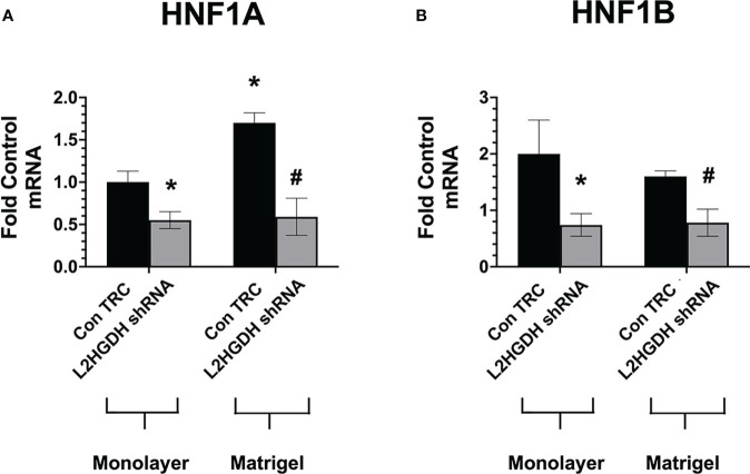
2-Hydroxyglutarate (2HG) overproducing tumors arise in a number of tissues, including the kidney. The tumorigenesis resulting from overproduced 2HG has been attributed to the ability of 2HG alter gene expression by inhibiting α-ketoglutarate (αKG)-dependent dioxygenases, including Ten-eleven-Translocation (TET) enzymes. Genes that regulate cellular differentiation are reportedly repressed, blocking differentiation of mesenchymal cells into myocytes, and adipocytes. In this report, the expression of the enzyme responsible for L2HG degradation, L-2HG dehydrogenase (L2HGDH), is knocked down, using lentiviral shRNA, as well as siRNA, in primary cultures of normal Renal Proximal Tubule (RPT) cells. The knockdown (KD) results in increased L-2HG levels, decreased demethylation of 5mC in genomic DNA, and increased methylation of H3 Histones. Consequences include reduced tubulogenesis by RPT cells in matrigel, and reduced expression of molecular markers of differentiation, including membrane transporters as well as HNF1α and HNF1β, which regulate their transcription. These results are consistent with the hypothesis that oncometabolite 2HG blocks RPT differentiation by altering the methylation status of chromatin in a manner that impedes the transcriptional events required for normal differentiation. Presumably, similar alterations are responsible for promoting the expansion of renal cancer stem-cells, increasing their propensity for malignant transformation.
2-羟戊二酸(2HG)在许多组织中产生肿瘤,包括肾脏。由于 2HG 的过度产生,通过抑制 α-酮戊二酸(αKG)依赖性双加氧酶,包括 Ten-eleven-Translocation(TET)酶,从而导致肿瘤发生。据报道,调节细胞分化的基因受到抑制,阻止间充质细胞分化为肌细胞和成脂细胞。在本报告中,使用慢病毒 shRNA 和 siRNA 敲低负责 L2HG 降解的酶,即 L-2HG 脱氢酶(L2HGDH),在正常肾近端小管(RPT)细胞的原代培养物中。敲低(KD)导致 L-2HG 水平升高、基因组 DNA 中 5mC 的去甲基化减少和 H3 组蛋白的甲基化增加。结果包括 RPT 细胞在基质胶中的小管形成减少,以及分化的分子标志物表达减少,包括膜转运体以及调节其转录的 HNF1α 和 HNF1β。这些结果与假设一致,即致癌代谢物 2HG 通过改变染色质的甲基化状态来阻止正常分化所需的转录事件,从而阻止 RPT 分化。推测类似的改变负责促进肾癌细胞干细胞的扩增,增加其恶性转化的倾向。