Gireadă Roxana, Socolov Demetra, Mihălceanu Elena, Lazăr Ioan Tudor, Luca Alexandru, Matasariu Roxana, Ursache Alexandra, Bujor Iuliana, Gireadă Tiberiu, Boiculese Vasile Lucian, Socolov Răzvan
Department of Obstetrics and Gynecology, University of Medicine and Pharmacy 'Gr. T. Popa', 700115 Iaşi, Romania.
Department of Obstetrics and Gynecology, Cuza Vodă Hospital, 700038 Iaşi, Romania.
Diagnostics (Basel). 2022 Aug 24;12(9):2053. doi: 10.3390/diagnostics12092053.
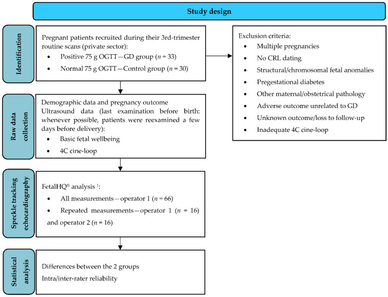
Background: The most commonly known cardiac effect of gestational diabetes mellitus (GD) in the fetus is hypertrophic cardiomyopathy, but recent studies show that it is preceded by subclinical cardiac dysfunction. This study aimed to assess the effect of GD on fetal cardiac geometry and contractility by two-dimensional speckle-tracking technology. Methods: We performed a prospective observational study that included 33 pregnant patients with GD and 30 healthy individuals. For all fetuses, a four-chamber 3 s cine-loop was recorded and analyzed with Fetal Heart Quantification (FetalHQ®), a novel proprietary speckle-tracking software. The following cardiac indices were calculated: global sphericity index (GSI), global longitudinal strain (GLS), fractional area change (FAC), and 24-segment end-diastolic diameter (EDD), fractional shortening (FS), and sphericity index (SI) for both ventricles. Demographic and cardiac differences between the two groups were analyzed, as well as intra-rater and inter-rater reliability. Results: There were significant changes in right ventricular FAC and FS for segments 4−24 in fetuses exposed to GD (−1 SD, p < 0.05). No significant differences were detected for GSI, GLS, EDD, or SI for either ventricle. Conclusions: Fetuses exposed to GD present impaired right ventricular contractility, especially in the mid and apical segments.
妊娠期糖尿病(GD)对胎儿最广为人知的心脏影响是肥厚型心肌病,但最近的研究表明,在此之前存在亚临床心脏功能障碍。本研究旨在通过二维斑点追踪技术评估GD对胎儿心脏几何形状和收缩功能的影响。方法:我们进行了一项前瞻性观察性研究,纳入了33例患有GD的孕妇和30名健康个体。对所有胎儿,记录一个四腔心3秒电影环,并使用一种新型的专有斑点追踪软件Fetal Heart Quantification(FetalHQ®)进行分析。计算以下心脏指标:整体球形指数(GSI)、整体纵向应变(GLS)、面积变化分数(FAC)以及两个心室的24节段舒张末期直径(EDD)、缩短分数(FS)和球形指数(SI)。分析了两组之间的人口统计学和心脏差异,以及评分者内和评分者间的可靠性。结果:暴露于GD的胎儿右心室4−24节段的FAC和FS有显著变化(−1标准差,p<0.05)。两个心室的GSI、GLS、EDD或SI均未检测到显著差异。结论:暴露于GD的胎儿右心室收缩功能受损,尤其是在中间段和心尖段。