• 文献检索
  • 文档翻译
  • 深度研究
  • 学术资讯
  • Suppr Zotero 插件Zotero 插件
  • 邀请有礼
  • 套餐&价格
  • 历史记录
应用&插件
Suppr Zotero 插件Zotero 插件浏览器插件Mac 客户端Windows 客户端微信小程序
定价
高级版会员购买积分包购买API积分包
服务
文献检索文档翻译深度研究API 文档MCP 服务
关于我们
关于 Suppr公司介绍联系我们用户协议隐私条款
关注我们

Suppr 超能文献

核心技术专利:CN118964589B侵权必究
粤ICP备2023148730 号-1Suppr @ 2026

文献检索

告别复杂PubMed语法,用中文像聊天一样搜索,搜遍4000万医学文献。AI智能推荐,让科研检索更轻松。

立即免费搜索

文件翻译

保留排版,准确专业,支持PDF/Word/PPT等文件格式,支持 12+语言互译。

免费翻译文档

深度研究

AI帮你快速写综述,25分钟生成高质量综述,智能提取关键信息,辅助科研写作。

立即免费体验

新型TrueVue系列三维超声心动图:揭示先天性心脏病的病理形态

Novel TrueVue series of 3D echocardiography: Revealing the pathological morphology of congenital heart disease.

作者信息

Sun Feifei, Sun Aijiao, Chen Yixin, Xiao Yangjie, Zhang Xintong, Qiao Wei, Tan Xueying, Liang Yanxiao, Li Dongyu, Yang Shu, Ren Weidong

机构信息

Department of Ultrasound, Shengjing Hospital of China Medical University, Shenyang, China.

Department of Cardiac Surgery, Shengjing Hospital of China Medical University, Shenyang, China.

出版信息

Front Physiol. 2022 Sep 6;13:1000007. doi: 10.3389/fphys.2022.1000007. eCollection 2022.

DOI:10.3389/fphys.2022.1000007
PMID:36148295
原文链接:https://pmc.ncbi.nlm.nih.gov/articles/PMC9485809/
Abstract

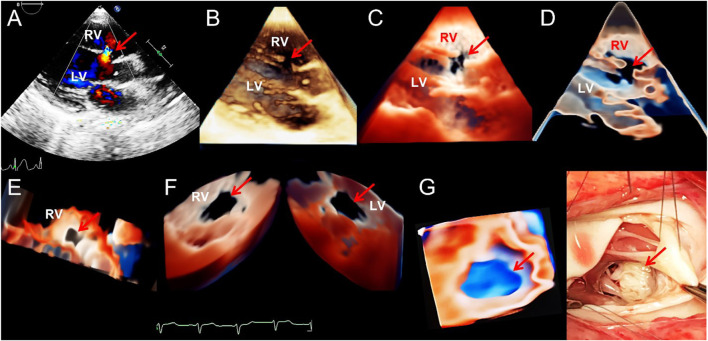
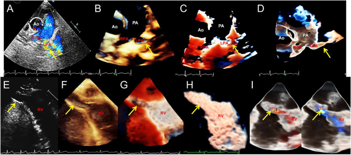
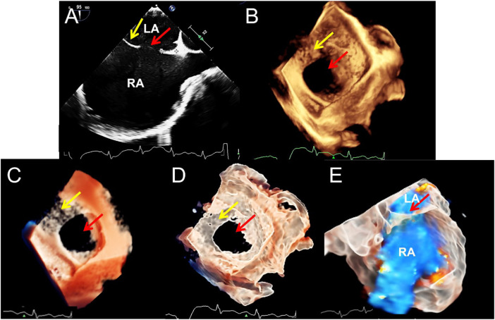
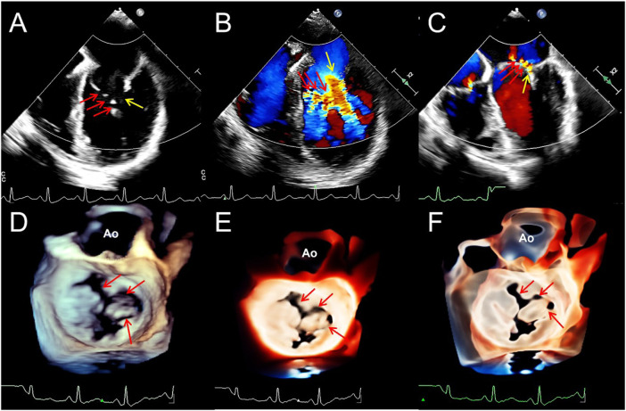
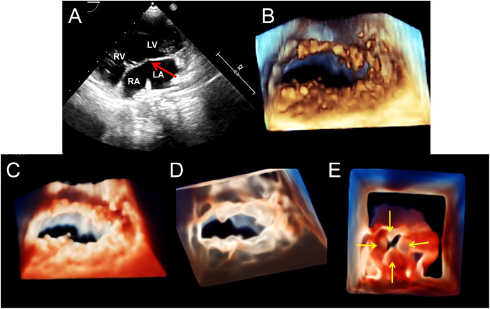
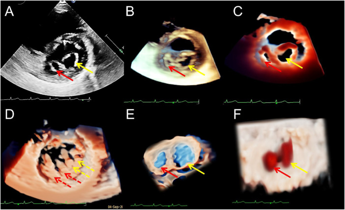
This study explored the advantages and limitations of novel series of three-dimensional (3D) echocardiographic techniques and summarized their application methods for congenital heart diseases (CHDs). Two-dimensional (2D), traditional 3D echocardiography, and TrueVue plus light and/or Glass novel 3D technologies were performed on 62 patients with CHD, and a clinical survey was designed to judge whether the novel 3D images were more helpful for understanding the cardiac condition and guide treatment than traditional 3D images. TrueVue increased the visual resolution and simulated the true texture of cardiac tissue, significantly improving the display ability of abnormal anatomical structures in CHDs. TrueVue Glass displayed the blood channel and the internal structure of cardiac cavity more intuitively, indicating a new observation aspect not shown by conventional echocardiography. The clinical survey results showed that the new 3D imaging methods effectively increased the diagnostic confidence of echocardiographers, enabled surgeons to better understand the details of lesions, promoted efficient communication, and improved the confidence of both doctors and patients in treatment. The combined application of TrueVue, TrueVue Light, and TrueVue Glass more closely simulated real anatomical features, showed more comprehensive and subtle blood flow in the lumen, not only increased the visual effect but also provided more useful diagnostic information, improved the accuracy of evaluation and treatment of CHD when compared to traditional imaging techniques, indicating that this combined application has significant clinical value.

摘要

本研究探讨了新型系列三维(3D)超声心动图技术的优势与局限性,并总结了其在先天性心脏病(CHD)中的应用方法。对62例CHD患者进行了二维(2D)、传统3D超声心动图以及TrueVue加光和/或Glass新型3D技术检查,并设计了一项临床调查,以判断新型3D图像在了解心脏状况和指导治疗方面是否比传统3D图像更有帮助。TrueVue提高了视觉分辨率,模拟了心脏组织的真实纹理,显著提高了CHD中异常解剖结构的显示能力。TrueVue Glass更直观地显示了血道和心腔内部结构,显示了传统超声心动图未显示的新观察视角。临床调查结果表明,新的3D成像方法有效提高了超声心动图医生的诊断信心,使外科医生能够更好地了解病变细节,促进了高效沟通,并提高了医生和患者对治疗的信心。TrueVue、TrueVue Light和TrueVue Glass的联合应用更紧密地模拟了真实解剖特征,显示管腔内血流更全面、细微,不仅增强了视觉效果,还提供了更多有用的诊断信息,与传统成像技术相比,提高了CHD评估和治疗的准确性,表明这种联合应用具有显著的临床价值。

https://cdn.ncbi.nlm.nih.gov/pmc/blobs/2356/9485809/51f03fdbfe8b/fphys-13-1000007-g006.jpg
https://cdn.ncbi.nlm.nih.gov/pmc/blobs/2356/9485809/ea9bcf79d285/fphys-13-1000007-g001.jpg
https://cdn.ncbi.nlm.nih.gov/pmc/blobs/2356/9485809/b10ee75e5286/fphys-13-1000007-g002.jpg
https://cdn.ncbi.nlm.nih.gov/pmc/blobs/2356/9485809/2dc87d894b5d/fphys-13-1000007-g003.jpg
https://cdn.ncbi.nlm.nih.gov/pmc/blobs/2356/9485809/31ee830363e7/fphys-13-1000007-g004.jpg
https://cdn.ncbi.nlm.nih.gov/pmc/blobs/2356/9485809/0ccfd9e516de/fphys-13-1000007-g005.jpg
https://cdn.ncbi.nlm.nih.gov/pmc/blobs/2356/9485809/51f03fdbfe8b/fphys-13-1000007-g006.jpg

相似文献

1
Novel TrueVue series of 3D echocardiography: Revealing the pathological morphology of congenital heart disease.新型TrueVue系列三维超声心动图:揭示先天性心脏病的病理形态
Front Physiol. 2022 Sep 6;13:1000007. doi: 10.3389/fphys.2022.1000007. eCollection 2022.
2
Real-time 3D echocardiographic transilluminated imaging combined with artificially intelligent left atrial appendage measurement for atrial fibrillation interventional procedures.实时三维超声心动图透照成像联合人工智能左心耳测量在房颤介入手术中的应用
Front Physiol. 2022 Nov 9;13:1043551. doi: 10.3389/fphys.2022.1043551. eCollection 2022.
3
Rare double orifice mitral valve malformation associated with bicuspid aortic valve in Turner syndrome: diagnosed by a series of novel three-dimensional echocardiography and literature review.特纳综合征相关罕见二尖瓣双孔畸形合并二叶式主动脉瓣:通过一系列新型三维超声心动图诊断并文献复习。
BMC Cardiovasc Disord. 2021 Aug 4;21(1):377. doi: 10.1186/s12872-021-02184-2.
4
Case report: Novel three-dimensional echocardiographic methods mapping aortic root pseudoaneurysm secondary to blood culture-negative endocarditis with bicuspid aortic valve involvement.病例报告:用于绘制继发于血培养阴性的心内膜炎且累及二叶式主动脉瓣的主动脉根部假性动脉瘤的新型三维超声心动图方法。
Front Cardiovasc Med. 2023 Mar 16;10:1138390. doi: 10.3389/fcvm.2023.1138390. eCollection 2023.
5
European Association of Cardiovascular Imaging/Cardiovascular Imaging Department of the Brazilian Society of Cardiology recommendations for the use of cardiac imaging to assess and follow patients after heart transplantation.欧洲心血管影像协会/巴西心脏病学会心血管影像学部关于心脏移植后评估和随访患者使用心脏影像的建议。
Eur Heart J Cardiovasc Imaging. 2015 Sep;16(9):919-48. doi: 10.1093/ehjci/jev139. Epub 2015 Jul 2.
6
Three-dimensional (3D) echocardiographic analysis of congenital heart disease in the fetus: comparison with cross-sectional (2D) fetal echocardiography.胎儿先天性心脏病的三维(3D)超声心动图分析:与横断面(2D)胎儿超声心动图的比较。
Ultrasound Obstet Gynecol. 2001 Jun;17(6):485-92. doi: 10.1046/j.1469-0705.2001.00429.x.
7
Dynamic three-dimensional display of common congenital cardiac defects from reconstruction of two-dimensional echocardiographic images.基于二维超声心动图图像重建的常见先天性心脏缺陷的动态三维显示
Zhonghua Min Guo Xiao Er Ke Yi Xue Hui Za Zhi. 1996 Jan-Feb;37(1):27-30.
8
A Novel Virtual Reality Medical Image Display System for Group Discussions of Congenital Heart Disease: Development and Usability Testing.一种用于先天性心脏病小组讨论的新型虚拟现实医学图像显示系统:开发与可用性测试
JMIR Cardio. 2020 Dec 8;4(1):e20633. doi: 10.2196/20633.
9
Three-dimensional printing of intracardiac defects from three-dimensional echocardiographic images: feasibility and relative accuracy.利用三维超声心动图图像进行心内缺损的三维打印:可行性与相对准确性
J Am Soc Echocardiogr. 2015 Apr;28(4):392-7. doi: 10.1016/j.echo.2014.12.016. Epub 2015 Feb 7.
10
Added value of three-/four-dimensional ultrasound in offline analysis and diagnosis of congenital heart disease.三维/四维超声在先天性心脏病离线分析诊断中的附加价值。
Ultrasound Obstet Gynecol. 2011 Apr;37(4):432-7. doi: 10.1002/uog.8867. Epub 2011 Mar 7.

引用本文的文献

1
Novel Techniques in Imaging Congenital Heart Disease: JACC Scientific Statement.先天性心脏病影像学的新方法:美国心脏病学会科学声明。
J Am Coll Cardiol. 2024 Jan 2;83(1):63-81. doi: 10.1016/j.jacc.2023.10.025.
2
Real-time 3D echocardiographic transilluminated imaging combined with artificially intelligent left atrial appendage measurement for atrial fibrillation interventional procedures.实时三维超声心动图透照成像联合人工智能左心耳测量在房颤介入手术中的应用
Front Physiol. 2022 Nov 9;13:1043551. doi: 10.3389/fphys.2022.1043551. eCollection 2022.

本文引用的文献

1
Prenatal diagnosis of fetal right ventricular diverticulum with massive pericardial effusion in one of monochorionic diamniotic twins: a case report with a favorable outcome following in utero pericardiocentesis.超声检查发现一例单绒毛膜双羊膜囊双胎之一胎儿右心室憩室伴大量心包积液:宫内心包穿刺术后结局良好的病例报告。
J Int Med Res. 2021 Jan;49(1):300060520986668. doi: 10.1177/0300060520986668.
2
Normal reference values for mechanical mitral prosthetic valve inner diameters and areas assessed by two-dimensional and real-time three-dimensional transesophageal echocardiography.二维及实时三维经食管超声心动图评估机械二尖瓣人工瓣环内径和面积的正常值。
Int J Cardiovasc Imaging. 2021 Feb;37(2):547-557. doi: 10.1007/s10554-020-02039-5. Epub 2020 Oct 3.
3
Improved Delineation of Cardiac Pathology Using a Novel Three-Dimensional Echocardiographic Tissue Transparency Tool.使用新型三维超声心动图组织透明度工具改善心脏病理学的描绘。
J Am Soc Echocardiogr. 2020 Nov;33(11):1316-1323. doi: 10.1016/j.echo.2020.08.005. Epub 2020 Sep 21.
4
Prenatal sonographic diagnosis of partial left pulmonary artery sling: A rare case report.产前超声诊断部分性左肺动脉吊带:罕见病例报告。
J Clin Ultrasound. 2021 Mar;49(3):257-261. doi: 10.1002/jcu.22913. Epub 2020 Sep 14.
5
Assessing the clinical significance of echocardiograms in determining treatment of patent ductus arteriosus in neonates.评估超声心动图在确定新生儿动脉导管未闭治疗中的临床意义。
J Neonatal Perinatal Med. 2020;13(3):345-350. doi: 10.3233/NPM-170122.
6
Stereoscopic Display Is Superior to Conventional Display for Three-Dimensional Echocardiography of Congenital Heart Anatomy.立体显示器优于传统显示器,用于先天性心脏病解剖的三维超声心动图。
J Am Soc Echocardiogr. 2020 Nov;33(11):1297-1305. doi: 10.1016/j.echo.2020.06.016. Epub 2020 Sep 9.
7
Heart transplantation at a single tertiary adult congenital heart disease centre: Too little, too late?单中心成人先天性心脏病心脏移植:做得太少,为时太晚?
Int J Cardiol. 2021 Jan 1;322:107-113. doi: 10.1016/j.ijcard.2020.08.047. Epub 2020 Aug 14.
8
4D flow cardiac magnetic resonance in children and adults with congenital heart disease: Clinical experience in a high volume center.先天性心脏病儿童和成人的4D流心脏磁共振成像:高容量中心的临床经验
Int J Cardiol. 2020 Dec 1;320:168-177. doi: 10.1016/j.ijcard.2020.07.021. Epub 2020 Jul 24.
9
Toward rational management of patent ductus arteriosus: ductal disease staging and first line paracetamol.朝向动脉导管未闭的合理管理:导管病变分期和一线扑热息痛。
J Matern Fetal Neonatal Med. 2021 Dec;34(23):3940-3945. doi: 10.1080/14767058.2019.1702949. Epub 2019 Dec 29.
10
Use of a photo-realism 3D rendering technique to enhance echocardiographic visualization of the anatomical details during beating-heart mitral valve repair.使用逼真的3D渲染技术增强心脏跳动时二尖瓣修复过程中解剖细节的超声心动图可视化。
Echocardiography. 2019 Nov;36(11):2090-2093. doi: 10.1111/echo.14515. Epub 2019 Nov 4.