Ai Rongshuang, Li Dingyi, Shi Luyao, Zhang Xiaonan, Ding Zhiqiang, Zhu Yiting, He Yujuan
Key Laboratory of Diagnostic Medicine (Ministry of Education), Department of Laboratory Medicine, Chongqing Medical University, Chongqing, China.
College of Stomatology, Chongqing Medical University, Chongqing, China.
Front Microbiol. 2022 Sep 8;13:875091. doi: 10.3389/fmicb.2022.875091. eCollection 2022.
To assess the contribution of polymicrobial disruption of host homeostasis to periodontitis progression in orthodontic wire ligation murine model.
Orthodontic wire rings were inserted between the first and second molars of mice for 18 days for the orthodontic wire ligation mouse model, and injection model and -LPS injection model were used as controls. Alveolar bone loss and periodontal inflammation were analyzed by micro-CT, histological staining and qRT-PCR. Further, pyrosequencing of 16S rRNA gene amplicon was used to analyze the development of oral microorganism dysbiosis in the mice.
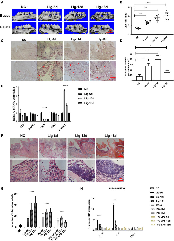
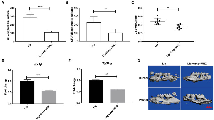
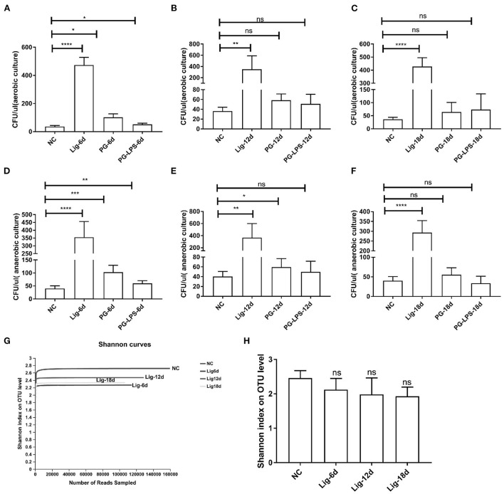
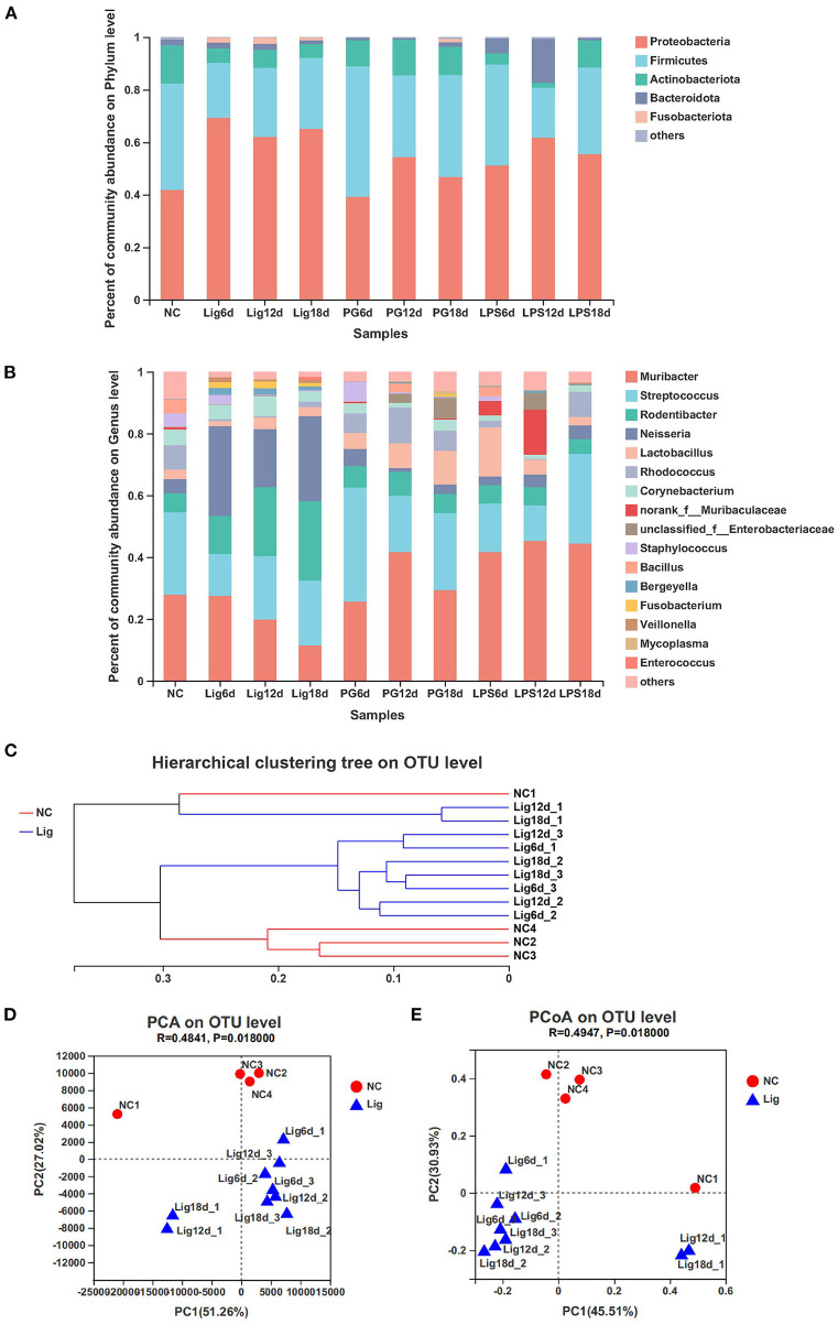
Micro-CT, TRAP staining and qRT-PCR showed that orthodontic wire ligation model led to more severe alveolar bone loss than and -LPS models.H&E staining and qRT-PCR demonstrated that stronger inflammatory response was induced by the orthodontic wire treatment compared to the other models. In addition, pyrosequencing of 16S rRNA gene amplicons revealed that the composition of oral microbiota presented a transition as the disease progressed and significant differences emerged in oral microbiota communities between orthodontic ligature mice and healthy controls. Furthermore, antibiotic treatment decreased both inflammation and alveolar bone loss in response to microbial community dysbiosis. However, no significant difference in bacterial community composition was observed in and -LPS models.
Orthodontic wire ligation drove oral microbial community transitions that mimicked polymicrobial communities characterized by polymicrobial synergy and dysbiosis. Our improved model is suitable for further study of pathogenesis of periodontitis and exploration of corresponding treatment strategies.
在正畸结扎小鼠模型中评估宿主稳态的多微生物破坏对牙周炎进展的作用。
将正畸结扎丝环插入小鼠第一和第二磨牙之间18天以建立正畸结扎小鼠模型,注射模型和脂多糖(LPS)注射模型作为对照。通过显微CT、组织学染色和定量逆转录聚合酶链反应(qRT-PCR)分析牙槽骨丧失和牙周炎症。此外,对16S rRNA基因扩增子进行焦磷酸测序,以分析小鼠口腔微生物群落失调的发展情况。
显微CT、抗酒石酸酸性磷酸酶(TRAP)染色和qRT-PCR显示,正畸结扎模型导致的牙槽骨丧失比注射模型和LPS模型更严重。苏木精-伊红(H&E)染色和qRT-PCR表明,与其他模型相比,正畸结扎处理诱导了更强的炎症反应。此外,16S rRNA基因扩增子的焦磷酸测序显示,随着疾病进展,口腔微生物群的组成发生了转变,正畸结扎小鼠与健康对照之间的口腔微生物群落出现了显著差异。此外,抗生素治疗减轻了因微生物群落失调引起的炎症和牙槽骨丧失。然而,在注射模型和LPS模型中未观察到细菌群落组成的显著差异。
正畸结扎促使口腔微生物群落转变,模拟了以多微生物协同作用和失调为特征的多微生物群落。我们改进的模型适用于进一步研究牙周炎的发病机制和探索相应的治疗策略。