Goldschleger Eye Institute, Sheba Medical Center, Tel Hashomer, Israel, Affiliated to the Sackler Faculty of Medicine, Tel Aviv University, Tel Aviv, Israel.
Indian J Ophthalmol. 2022 Oct;70(10):3562-3568. doi: 10.4103/ijo.IJO_84_22.
To examine the differences in the peripapillary vascular parameters and foveal-avascular-zone (FAZ) vascularity parameters between primary open-angle-glaucoma (POAG) patients versus exfoliation-glaucoma (XFG) patients versus healthy subjects.
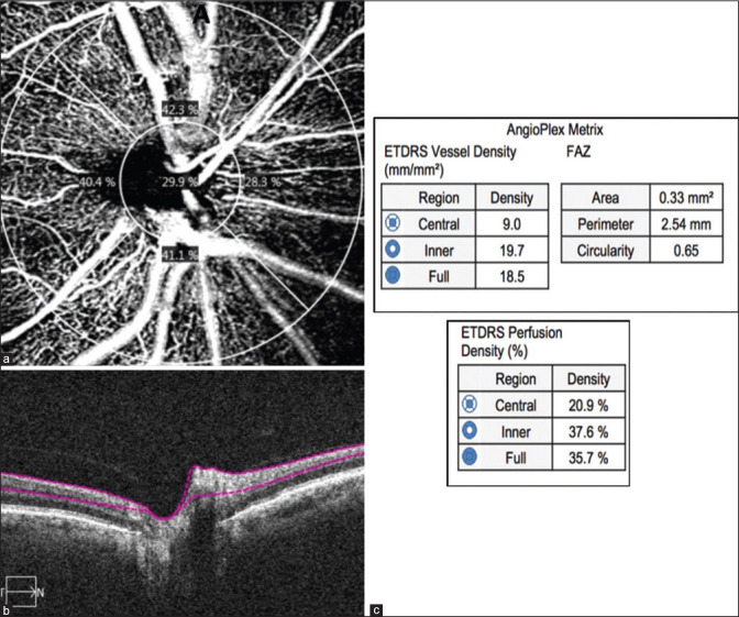
This is cross-sectional study and a comparative clinical study. POAG and XFG patients and healthy subjects underwent a comprehensive ophthalmic examination, including visual field optical coherence tomography (OCT) and OCT angiography (OCTA) of the optic disc and FAZ. Differences in peripapillary vessel density (VD), perfusion density (PD), and FAZ area and circularity were examined between all groups, as well as correlations between clinical parameters and vascularity parameters for each glaucoma group.
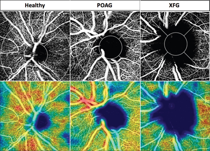
A total of 109 subjects (one eye for each patient) were analyzed, including 45 with POAG, 30 with XFG, and 34 controls. The average peripapillary VDs were the lowest among the XFG patients and the highest among the controls (P < 0.05, ANOVA). The average peripapillary PD of the central ring was the lowest in the XFG group and the highest in the control group (P = 0.02, ANOVA). A significant negative correlation was found between the average peripapillary VDs and PDs of the inner ring and full ring and disease severity of the POAG patients. There was a significant positive correlation between the average peripapillary PDs of the central rings and full ring and the central macular thickness of the XFG patients (P < 0.01 and P < 0.04, respectively, Pearson correlation).
The peripapillary vascular parameters of the POAG and XFG patients were lower compared to those of normal participants. A correlation between clinical characteristics of POAG and XFG patients and PD was found. This may hint to a vascular mechanism in glaucoma either primary or secondary to intra-ocular pressure/OAG damage.
研究原发性开角型青光眼(POAG)患者、剥脱性青光眼(XFG)患者与健康受试者之间视盘旁血管参数和中心凹无血管区(FAZ)血管密度参数的差异。
这是一项横断面研究和对比临床研究。POAG 和 XFG 患者以及健康受试者接受了全面的眼科检查,包括视野光学相干断层扫描(OCT)和视盘及 FAZ 的 OCT 血管造影(OCTA)。检查了所有组之间的视盘旁血管密度(VD)、灌注密度(PD)、FAZ 面积和圆度的差异,以及每个青光眼组的临床参数与血管密度参数之间的相关性。
共分析了 109 名受试者(每位患者一只眼),其中 45 名患有 POAG,30 名患有 XFG,34 名作为对照组。XFG 患者的平均视盘旁 VD 最低,对照组最高(P<0.05,方差分析)。XFG 组中央环的平均视盘旁 PD 最低,对照组最高(P=0.02,方差分析)。POAG 患者的平均视盘旁内环和全环 VD 和 PD 与疾病严重程度呈显著负相关。XFG 患者的中央环和全环平均视盘旁 PD 与中央黄斑厚度呈显著正相关(P<0.01 和 P<0.04,Pearson 相关)。
POAG 和 XFG 患者的视盘旁血管参数低于正常受试者。POAG 和 XFG 患者的临床特征与 PD 之间存在相关性。这可能提示原发性或继发性眼压/开角型青光眼损害的青光眼存在血管机制。