• 文献检索
  • 文档翻译
  • 深度研究
  • 学术资讯
  • Suppr Zotero 插件Zotero 插件
  • 邀请有礼
  • 套餐&价格
  • 历史记录
应用&插件
Suppr Zotero 插件Zotero 插件浏览器插件Mac 客户端Windows 客户端微信小程序
定价
高级版会员购买积分包购买API积分包
服务
文献检索文档翻译深度研究API 文档MCP 服务
关于我们
关于 Suppr公司介绍联系我们用户协议隐私条款
关注我们

Suppr 超能文献

核心技术专利:CN118964589B侵权必究
粤ICP备2023148730 号-1Suppr @ 2026

文献检索

告别复杂PubMed语法,用中文像聊天一样搜索,搜遍4000万医学文献。AI智能推荐,让科研检索更轻松。

立即免费搜索

文件翻译

保留排版,准确专业,支持PDF/Word/PPT等文件格式,支持 12+语言互译。

免费翻译文档

深度研究

AI帮你快速写综述,25分钟生成高质量综述,智能提取关键信息,辅助科研写作。

立即免费体验

利用影像学区分多发性硬化与水通道蛋白 4 相关视神经脊髓炎谱系疾病和髓鞘少突胶质细胞糖蛋白抗体病。

Differentiating Multiple Sclerosis From AQP4-Neuromyelitis Optica Spectrum Disorder and MOG-Antibody Disease With Imaging.

机构信息

From the Department of Neuroinflammation (R.C., F.P.C., C.T., A.B., W.B., F.D.A., I.D.L.P., F.G., L.H., L.M., A.T., M.Y., A.T.T., Y.H.R.C.P.C.H., F.B., O.C.), Queen Square MS Centre, UCL Queen Square Institute of Neurology, Faculty of Brain Science, University College of London; Department of Medicine (R.C.), Surgery and Neuroscience, University of Siena, Italy; Department of Medical Physics and Biomedical Engineering (F.P.C., B.K., F.B.), Centre for Medical Imaging Computing, University College of London; Universitat Oberta de Catalunya (F.P.C.), Barcelona, Spain; MS Centre of Catalonia (Cemcat) (C.T.), Vall d'Hebron Institute of Research, Spain; Radiomics Group (F.G.), Vall d'Hebron Institute of Oncology, Vall d'Hebron Barcelona Barcelona, Spain; Department of Biomedical Imaging and Image Guided Therapy (L.H.), Medical University of Vienna, Austria; NMO Clinical Service at the Walton Centre (A.J.), Liverpool, United Kingdom; Division of Multiple Sclerosis and Autoimmune Neurology (A.J.), Neurological Institute, Cleveland Clinic Abu Dhabi, United Arab Emirates; Division of Brain Sciences (R.S.N.), Department of Medicine, Imperial College London; National Institute for Health Research (NIHR) (A.T., F.B., O.C.), University College London Hospitals (UCLH), Biomedical Research Centre; and Department of Radiology and Nuclear Medicine (F.B.), Amsterdam University Medical Centre, the Netherlands.

出版信息

Neurology. 2023 Jan 17;100(3):e308-e323. doi: 10.1212/WNL.0000000000201465. Epub 2022 Oct 3.

DOI:10.1212/WNL.0000000000201465
PMID:36192175
原文链接:https://pmc.ncbi.nlm.nih.gov/articles/PMC9869760/
Abstract

BACKGROUND AND OBJECTIVES

Relapsing-remitting multiple sclerosis (RRMS), aquaporin-4 antibody-positive neuromyelitis optica spectrum disorder (AQP4-NMOSD), and myelin oligodendrocyte glycoprotein antibody-associated disease (MOGAD) may have overlapping clinical features. There is an unmet need for imaging markers that differentiate between them when serologic testing is unavailable or ambiguous. We assessed whether imaging characteristics typical of MS discriminate RRMS from AQP4-NMOSD and MOGAD, alone and in combination.

METHODS

Adult, nonacute patients with RRMS, APQ4-NMOSD, and MOGAD and healthy controls were prospectively recruited at the National Hospital for Neurology and Neurosurgery (London, United Kingdom) and the Walton Centre (Liverpool, United Kingdom) between 2014 and 2019. They underwent conventional and advanced brain, cord, and optic nerve MRI and optical coherence tomography (OCT).

RESULTS

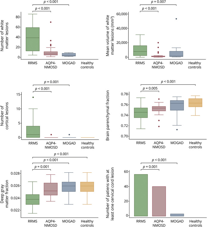
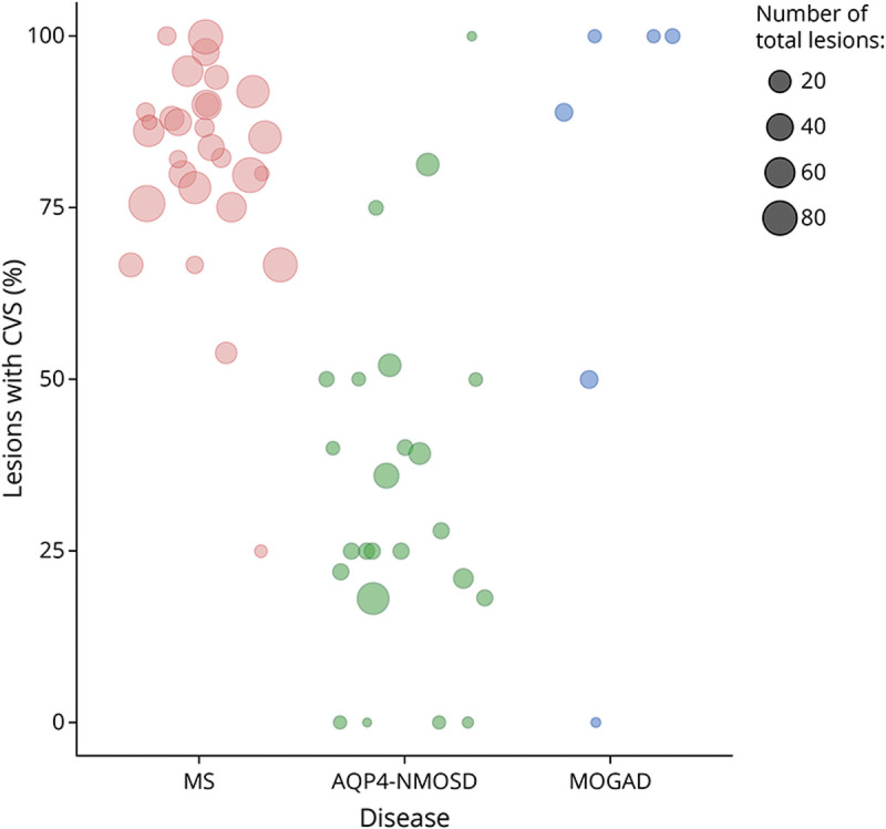
A total of 91 consecutive patients (31 RRMS, 30 APQ4-NMOSD, and 30 MOGAD) and 34 healthy controls were recruited. The most accurate measures differentiating RRMS from AQP4-NMOSD were the proportion of lesions with the central vein sign (CVS) (84% vs 33%, accuracy/specificity/sensitivity: 91/88/93%, < 0.001), followed by cortical lesions (median: 2 [range: 1-14] vs 1 [0-1], accuracy/specificity/sensitivity: 84/90/77%, = 0.002) and white matter lesions (mean: 39.07 [±25.8] vs 9.5 [±14], accuracy/specificity/sensitivity: 78/84/73%, = 0.001). The combination of higher proportion of CVS, cortical lesions, and optic nerve magnetization transfer ratio reached the highest accuracy in distinguishing RRMS from AQP4-NMOSD (accuracy/specificity/sensitivity: 95/92/97%, < 0.001). The most accurate measures favoring RRMS over MOGAD were white matter lesions (39.07 [±25.8] vs 1 [±2.3], accuracy/specificity/sensitivity: 94/94/93%, = 0.006), followed by cortical lesions (2 [1-14] vs 1 [0-1], accuracy/specificity/sensitivity: 84/97/71%, = 0.004), and retinal nerve fiber layer thickness (RNFL) (mean: 87.54 [±13.83] vs 75.54 [±20.33], accuracy/specificity/sensitivity: 80/79/81%, = 0.009). Higher cortical lesion number combined with higher RNFL thickness best differentiated RRMS from MOGAD (accuracy/specificity/sensitivity: 84/92/77%, < 0.001).

DISCUSSION

Cortical lesions, CVS, and optic nerve markers achieve a high accuracy in distinguishing RRMS from APQ4-NMOSD and MOGAD. This information may be useful in clinical practice, especially outside the acute phase and when serologic testing is ambiguous or not promptly available.

CLASSIFICATION OF EVIDENCE

This study provides Class II evidence that selected conventional and advanced brain, cord, and optic nerve MRI and OCT markers distinguish adult patients with RRMS from AQP4-NMOSD and MOGAD.

摘要

背景与目的

复发缓解型多发性硬化症(RRMS)、水通道蛋白 4 抗体阳性视神经脊髓炎谱系疾病(AQP4-NMOSD)和髓鞘少突胶质细胞糖蛋白抗体相关性疾病(MOGAD)可能具有重叠的临床特征。在血清学检测不可用或不明确时,需要影像学标志物来区分它们,但目前这一需求尚未得到满足。我们评估了典型的 MS 影像学特征是否可以单独或组合区分 RRMS 与 AQP4-NMOSD 和 MOGAD。

方法

2014 年至 2019 年,在英国国家神经学和神经外科学院(伦敦)和沃尔顿中心(利物浦)前瞻性招募了成人非急性 RRMS、AQP4-NMOSD 和 MOGAD 患者以及健康对照者。他们接受了常规和高级脑部、脊髓和视神经 MRI 及光学相干断层扫描(OCT)检查。

结果

共纳入 91 例连续患者(31 例 RRMS、30 例 AQP4-NMOSD 和 30 例 MOGAD)和 34 例健康对照者。区分 RRMS 与 AQP4-NMOSD 最准确的指标是伴有中央静脉征(CVS)的病灶比例(84%比 33%,准确性/特异性/敏感性:91/88/93%,<0.001),其次是皮质病灶(中位数:2[范围:1-14]比 1[0-1],准确性/特异性/敏感性:84/90/77%,=0.002)和白质病灶(均值:39.07[±25.8]比 9.5[±14],准确性/特异性/敏感性:78/84/73%,=0.001)。CVS 比例较高、皮质病灶和视神经磁化传递比的组合在区分 RRMS 与 AQP4-NMOSD 方面达到了最高的准确性(准确性/特异性/敏感性:95/92/97%,<0.001)。有利于 RRMS 的最准确指标是白质病灶(39.07[±25.8]比 1[±2.3],准确性/特异性/敏感性:94/94/93%,=0.006),其次是皮质病灶(2[1-14]比 1[0-1],准确性/特异性/敏感性:84/97/71%,=0.004)和视网膜神经纤维层厚度(RNFL)(均值:87.54[±13.83]比 75.54[±20.33],准确性/特异性/敏感性:80/79/81%,=0.009)。皮质病灶数量较高与 RNFL 厚度较高相结合可最佳地区分 RRMS 与 MOGAD(准确性/特异性/敏感性:84/92/77%,<0.001)。

讨论

皮质病灶、CVS 和视神经标志物在区分 RRMS 与 AQP4-NMOSD 和 MOGAD 方面具有很高的准确性。这些信息在临床实践中可能有用,尤其是在急性期之外,当血清学检测不明确或不能及时获得时。

证据分类

本研究提供了 II 级证据,表明选定的常规和高级脑、脊髓和视神经 MRI 和 OCT 标志物可区分成人 RRMS 患者与 AQP4-NMOSD 和 MOGAD。

https://cdn.ncbi.nlm.nih.gov/pmc/blobs/f339/9869760/e198a214b671/WNL-2022-201326f3.jpg
https://cdn.ncbi.nlm.nih.gov/pmc/blobs/f339/9869760/32b69d1a7699/WNL-2022-201326f1.jpg
https://cdn.ncbi.nlm.nih.gov/pmc/blobs/f339/9869760/07fa3a612dbd/WNL-2022-201326f2.jpg
https://cdn.ncbi.nlm.nih.gov/pmc/blobs/f339/9869760/e198a214b671/WNL-2022-201326f3.jpg
https://cdn.ncbi.nlm.nih.gov/pmc/blobs/f339/9869760/32b69d1a7699/WNL-2022-201326f1.jpg
https://cdn.ncbi.nlm.nih.gov/pmc/blobs/f339/9869760/07fa3a612dbd/WNL-2022-201326f2.jpg
https://cdn.ncbi.nlm.nih.gov/pmc/blobs/f339/9869760/e198a214b671/WNL-2022-201326f3.jpg

相似文献

1
Differentiating Multiple Sclerosis From AQP4-Neuromyelitis Optica Spectrum Disorder and MOG-Antibody Disease With Imaging.利用影像学区分多发性硬化与水通道蛋白 4 相关视神经脊髓炎谱系疾病和髓鞘少突胶质细胞糖蛋白抗体病。
Neurology. 2023 Jan 17;100(3):e308-e323. doi: 10.1212/WNL.0000000000201465. Epub 2022 Oct 3.
2
Grey Matter Atrophy and its Relationship with White Matter Lesions in Patients with Myelin Oligodendrocyte Glycoprotein Antibody-associated Disease, Aquaporin-4 Antibody-Positive Neuromyelitis Optica Spectrum Disorder, and Multiple Sclerosis.少突胶质细胞糖蛋白抗体相关疾病、水通道蛋白 4 抗体阳性视神经脊髓炎谱系疾病和多发性硬化症患者的灰质萎缩及其与白质病变的关系。
Ann Neurol. 2024 Aug;96(2):276-288. doi: 10.1002/ana.26951. Epub 2024 May 23.
3
Optic chiasm involvement in multiple sclerosis, aquaporin-4 antibody-positive neuromyelitis optica spectrum disorder and myelin oligodendrocyte glycoprotein-associated disease.多发性硬化症、水通道蛋白 4 抗体阳性视神经脊髓炎谱系疾病和髓鞘少突胶质细胞糖蛋白相关疾病中的视交叉受累。
Mult Scler. 2024 May;30(6):674-686. doi: 10.1177/13524585241240420. Epub 2024 Apr 22.
4
Central vein sign and other radiographic features distinguishing myelin oligodendrocyte glycoprotein antibody disease from multiple sclerosis and aquaporin-4 antibody-positive neuromyelitis optica.中央静脉征及其他影像学特征鉴别髓鞘少突胶质细胞糖蛋白抗体病与多发性硬化和水通道蛋白 4 抗体阳性视神经脊髓炎。
Mult Scler. 2022 Jan;28(1):49-60. doi: 10.1177/13524585211007086. Epub 2021 Apr 19.
5
Modified models to distinguish central nervous system demyelinating diseases with brain lesions.用于区分有脑部病变的中枢神经系统脱髓鞘疾病的改良模型。
Mult Scler Relat Disord. 2021 Jul;52:102965. doi: 10.1016/j.msard.2021.102965. Epub 2021 Apr 17.
6
Progression independent of relapses in aquaporin4-IgG-seropositive neuromyelitis optica spectrum disorder, myelin oligodendrocyte glycoprotein antibody-associated disease, and multiple sclerosis.AQP4-IgG 阳性视神经脊髓炎谱系疾病、髓鞘少突胶质细胞糖蛋白抗体相关疾病和多发性硬化症中与复发无关的进展。
Mult Scler Relat Disord. 2023 Dec;80:105093. doi: 10.1016/j.msard.2023.105093. Epub 2023 Oct 20.
7
Contrasting the brain imaging features of MOG-antibody disease, with AQP4-antibody NMOSD and multiple sclerosis.对比 MO G 抗体病、AQP4 抗体 NMOSD 和多发性硬化症的脑影像学特征。
Mult Scler. 2022 Feb;28(2):217-227. doi: 10.1177/13524585211018987. Epub 2021 May 28.
8
Clinical and MRI measures to identify non-acute MOG-antibody disease in adults.用于识别成人非急性 MOG 抗体病的临床和 MRI 指标。
Brain. 2023 Jun 1;146(6):2489-2501. doi: 10.1093/brain/awac480.
9
Serum Neurofilament Light and GFAP Are Associated With Disease Severity in Inflammatory Disorders With Aquaporin-4 or Myelin Oligodendrocyte Glycoprotein Antibodies.血清神经丝轻链和 GFAP 与水通道蛋白 4 或髓鞘少突胶质细胞糖蛋白抗体阳性的炎症性疾病的严重程度相关。
Front Immunol. 2021 Mar 16;12:647618. doi: 10.3389/fimmu.2021.647618. eCollection 2021.
10
Tumefactive Demyelination in MOG Ab-Associated Disease, Multiple Sclerosis, and AQP-4-IgG-Positive Neuromyelitis Optica Spectrum Disorder.MOG 抗体相关性疾病、多发性硬化症和 AQP-4-IgG 阳性视神经脊髓炎谱系疾病中的肿块样脱髓鞘病变。
Neurology. 2023 Mar 28;100(13):e1418-e1432. doi: 10.1212/WNL.0000000000206820. Epub 2023 Jan 23.

引用本文的文献

1
Deep Learning Modeling to Differentiate Multiple Sclerosis From MOG Antibody-Associated Disease.用于区分多发性硬化症与MOG抗体相关疾病的深度学习建模
Neurology. 2025 Sep 23;105(6):e214075. doi: 10.1212/WNL.0000000000214075. Epub 2025 Sep 4.
2
A20 (TNFAIP3) Distinguishes Attack From Remission in Pediatric Patients With Monophasic MOGAD.A20(TNFAIP3)可区分单相性MOGAD儿科患者的发作期与缓解期。
Neurol Neuroimmunol Neuroinflamm. 2025 Sep;12(5):e200452. doi: 10.1212/NXI.0000000000200452. Epub 2025 Aug 18.
3
Alterations in peripheral blood immune cell profiles of neuromyelitis optica spectrum disorder across different phases and after B cell depletion therapy.

本文引用的文献

1
Myelin-oligodendrocyte glycoprotein antibody-associated disease.髓鞘少突胶质细胞糖蛋白抗体相关性疾病。
Lancet Neurol. 2021 Sep;20(9):762-772. doi: 10.1016/S1474-4422(21)00218-0.
2
Comparison of MRI Lesion Evolution in Different Central Nervous System Demyelinating Disorders.不同中枢神经系统脱髓鞘疾病的 MRI 病变演变比较。
Neurology. 2021 Sep 14;97(11):e1097-e1109. doi: 10.1212/WNL.0000000000012467. Epub 2021 Jul 14.
3
Contrasting the brain imaging features of MOG-antibody disease, with AQP4-antibody NMOSD and multiple sclerosis.
视神经脊髓炎谱系障碍在不同阶段及B细胞清除治疗后的外周血免疫细胞谱变化。
Front Immunol. 2025 Jun 11;16:1556259. doi: 10.3389/fimmu.2025.1556259. eCollection 2025.
4
A narrative review of the economic burden of myelin oligodendrocyte glycoprotein antibody-associated disease and analogous conditions.髓鞘少突胶质细胞糖蛋白抗体相关疾病及类似病症经济负担的叙述性综述。
Front Neurol. 2025 May 30;16:1506465. doi: 10.3389/fneur.2025.1506465. eCollection 2025.
5
Comorbidities Are Associated With Unfavorable Outcome in Aquaporin-4 Antibody Positive Neuromyelitis Optica Spectrum Disorders and Myelin Oligodendrocyte Glycoprotein Antibody-Associated Disease: Exploratory Study From the CROCTINO Cohort.合并症与水通道蛋白4抗体阳性视神经脊髓炎谱系障碍和髓鞘少突胶质细胞糖蛋白抗体相关疾病的不良预后相关:来自CROCTINO队列的探索性研究
Eur J Neurol. 2025 Jun;32(6):e70214. doi: 10.1111/ene.70214.
6
Brain Characteristics in Patients With Myelin Oligodendrocyte Glycoprotein Antibody-Associated Disorder by 7.0 Tesla MRI.7.0特斯拉磁共振成像对髓鞘少突胶质细胞糖蛋白抗体相关疾病患者脑特征的研究
Ann Clin Transl Neurol. 2025 Jul;12(7):1431-1441. doi: 10.1002/acn3.70080. Epub 2025 May 22.
7
Clinical integration of brain and cord MRI features improves differential diagnosis of multiple sclerosis.脑和脊髓MRI特征的临床整合改善了多发性硬化的鉴别诊断。
J Neurol. 2025 May 11;272(6):388. doi: 10.1007/s00415-025-13124-x.
8
A Case of Refractory Hiccups and Numbness as the Initial Presentation of Multiple Sclerosis.一例以难治性呃逆和麻木为首发表现的多发性硬化症
Cureus. 2025 Apr 5;17(4):e81774. doi: 10.7759/cureus.81774. eCollection 2025 Apr.
9
Brain Volume Measures in Adults with MOG-Antibody-Associated Disease: A Longitudinal Multicenter Study.成人MOG抗体相关疾病的脑容量测量:一项纵向多中心研究。
J Clin Med. 2025 Apr 3;14(7):2445. doi: 10.3390/jcm14072445.
10
The Kappa Free Light Chains Index and Central Vein Sign: Two New Biomarkers for Multiple Sclerosis Diagnosis.游离κ轻链指数与中央静脉征:多发性硬化诊断的两种新生物标志物。
Neurol Ther. 2025 Jun;14(3):711-731. doi: 10.1007/s40120-025-00737-7. Epub 2025 Apr 6.
对比 MO G 抗体病、AQP4 抗体 NMOSD 和多发性硬化症的脑影像学特征。
Mult Scler. 2022 Feb;28(2):217-227. doi: 10.1177/13524585211018987. Epub 2021 May 28.
4
Positive Predictive Value of Myelin Oligodendrocyte Glycoprotein Autoantibody Testing.髓鞘少突胶质细胞糖蛋白自身抗体检测的阳性预测值。
JAMA Neurol. 2021 Jun 1;78(6):741-746. doi: 10.1001/jamaneurol.2021.0912.
5
Central vein sign and other radiographic features distinguishing myelin oligodendrocyte glycoprotein antibody disease from multiple sclerosis and aquaporin-4 antibody-positive neuromyelitis optica.中央静脉征及其他影像学特征鉴别髓鞘少突胶质细胞糖蛋白抗体病与多发性硬化和水通道蛋白 4 抗体阳性视神经脊髓炎。
Mult Scler. 2022 Jan;28(1):49-60. doi: 10.1177/13524585211007086. Epub 2021 Apr 19.
6
Brain structural alterations in MOG antibody diseases: a comparative study with AQP4 seropositive NMOSD and MS.MOG 抗体病的脑结构改变:与 AQP4 阳性 NMOSD 和 MS 的对比研究。
J Neurol Neurosurg Psychiatry. 2021 Jul;92(7):709-716. doi: 10.1136/jnnp-2020-324826. Epub 2021 Mar 9.
7
Different patterns of brainstem and cerebellar MRI abnormalities in demyelinating disorders with MOG and aquaporin-4 antibodies.伴有髓鞘少突胶质细胞糖蛋白(MOG)和水通道蛋白4抗体的脱髓鞘疾病中脑干和小脑MRI异常的不同模式
J Neurol Neurosurg Psychiatry. 2021 Feb 8. doi: 10.1136/jnnp-2020-325503.
8
Quantitative spinal cord MRI in MOG-antibody disease, neuromyelitis optica and multiple sclerosis.定量脊髓 MRI 在 MOG 抗体病、视神经脊髓炎和多发性硬化中的应用。
Brain. 2021 Feb 12;144(1):198-212. doi: 10.1093/brain/awaa347.
9
AQP4-IgG and MOG-IgG Related Optic Neuritis-Prevalence, Optical Coherence Tomography Findings, and Visual Outcomes: A Systematic Review and Meta-Analysis.水通道蛋白4抗体(AQP4-IgG)和髓鞘少突胶质细胞糖蛋白抗体(MOG-IgG)相关视神经炎——患病率、光学相干断层扫描结果及视觉预后:一项系统评价和荟萃分析
Front Neurol. 2020 Oct 8;11:540156. doi: 10.3389/fneur.2020.540156. eCollection 2020.
10
Detection of MOG-IgG by cell-based assay: moving from discovery to clinical practice.基于细胞的检测方法检测 MOG-IgG:从发现到临床实践。
Neurol Sci. 2021 Jan;42(1):73-80. doi: 10.1007/s10072-020-04828-1. Epub 2020 Oct 15.