• 文献检索
  • 文档翻译
  • 深度研究
  • 学术资讯
  • Suppr Zotero 插件Zotero 插件
  • 邀请有礼
  • 套餐&价格
  • 历史记录
应用&插件
Suppr Zotero 插件Zotero 插件浏览器插件Mac 客户端Windows 客户端微信小程序
定价
高级版会员购买积分包购买API积分包
服务
文献检索文档翻译深度研究API 文档MCP 服务
关于我们
关于 Suppr公司介绍联系我们用户协议隐私条款
关注我们

Suppr 超能文献

核心技术专利:CN118964589B侵权必究
粤ICP备2023148730 号-1Suppr @ 2026

文献检索

告别复杂PubMed语法,用中文像聊天一样搜索,搜遍4000万医学文献。AI智能推荐,让科研检索更轻松。

立即免费搜索

文件翻译

保留排版,准确专业,支持PDF/Word/PPT等文件格式,支持 12+语言互译。

免费翻译文档

深度研究

AI帮你快速写综述,25分钟生成高质量综述,智能提取关键信息,辅助科研写作。

立即免费体验

标准弥散加权磁共振成像在巨细胞动脉炎缺血性视神经病变中的应用。

Utility of standard diffusion-weighted magnetic resonance imaging for the identification of ischemic optic neuropathy in giant cell arteritis.

机构信息

Department of Neurology, Charité - Universitätsmedizin Berlin, Corporate Member of Freie Universität Berlin and Humboldt Universität zu Berlin, Berlin, Germany.

Institute of Neuroradiology, Charité - Universitätsmedizin Berlin, Corporate Member of Freie Universität Berlin and Humboldt Universität zu Berlin, Berlin, Germany.

出版信息

Sci Rep. 2022 Oct 3;12(1):16553. doi: 10.1038/s41598-022-20916-y.

DOI:10.1038/s41598-022-20916-y
PMID:36192437
原文链接:https://pmc.ncbi.nlm.nih.gov/articles/PMC9530116/
Abstract

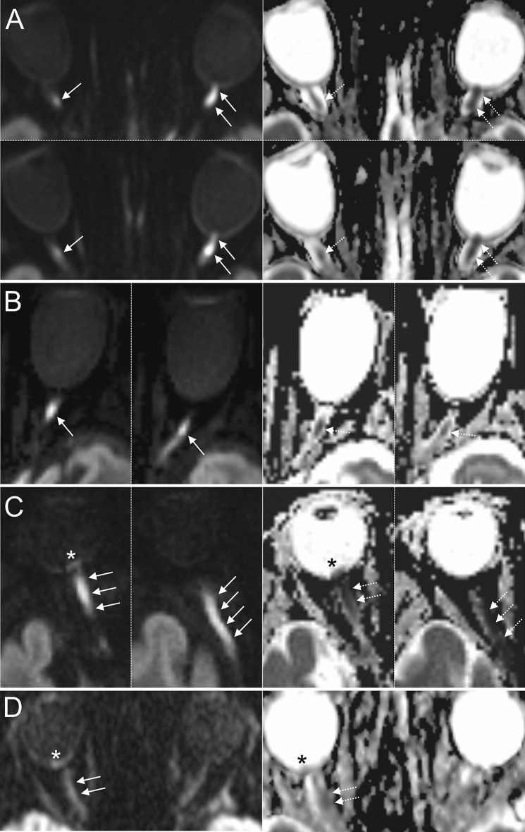
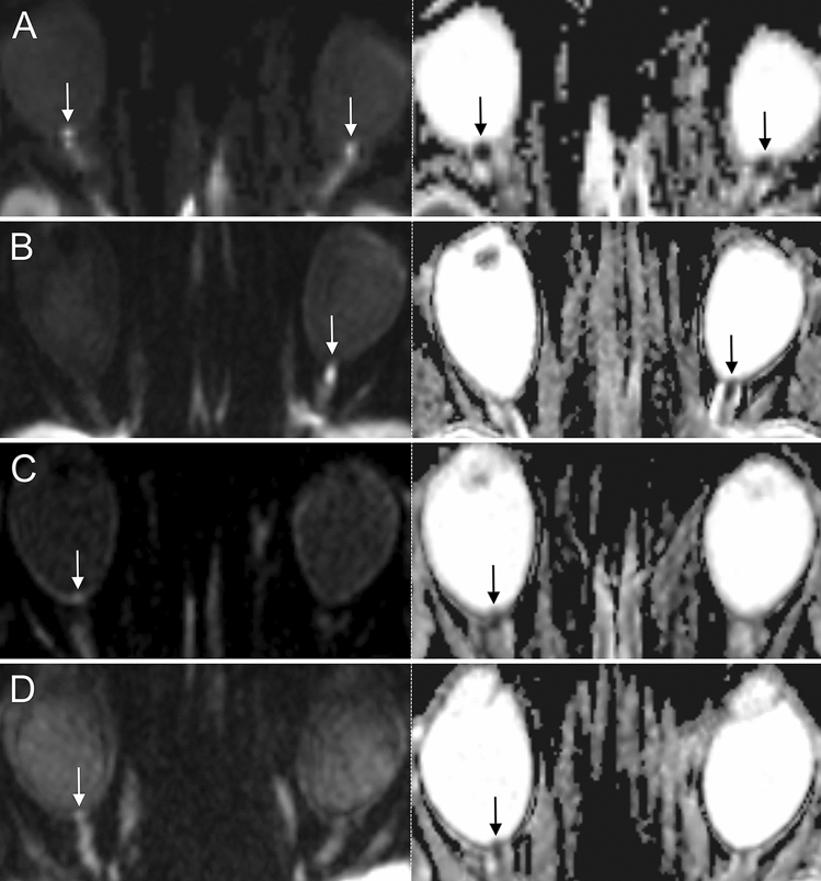
This study assessed diffusion abnormalities of the optic nerve (ON) in giant cell arteritis (GCA) patients with acute onset of visual impairment (VI) using diffusion-weighted magnetic resonance imaging (DWI). DWI scans of GCA patients with acute VI were evaluated in a case-control study. Two blinded neuroradiologists assessed randomized DWI scans of GCA and controls for ON restricted diffusion. Statistical quality criteria and inter-rater reliability (IRR) were calculated. DWI findings were compared to ophthalmological assessments. 35 GCA patients (76.2 ± 6.4 years; 37 scans) and 35 controls (75.7 ± 7.6 years; 38 scans) were included. ON restricted diffusion was detected in 81.1% (Reader 1) of GCA scans. Localization of ON restricted diffusion was at the optic nerve head in 80.6%, intraorbital in 11.1% and affecting both segments in 8.3%. DWI discerned affected from unaffected ON with a sensitivity, specificity, positive and negative predictive value of 87%/99%/96%/96%. IRR for ON restricted diffusion was κ = 0.72 (95% CI 0.59-0.86). DWI findings challenged ophthalmologic diagnoses in 4 cases (11.4%). DWI visualizes anterior and posterior ON ischemia in GCA patients with high sensitivity and specificity, as well as substantial IRR. DWI may complement the ophthalmological assessment in patients with acute VI.

摘要

本研究通过扩散加权磁共振成像(DWI)评估了急性视力障碍(VI)的巨细胞动脉炎(GCA)患者视神经(ON)的扩散异常。在一项病例对照研究中,对急性 VI 的 GCA 患者的 DWI 扫描进行了评估。两位盲法神经放射科医生对 GCA 和对照组的随机 DWI 扫描进行了 ON 受限扩散评估。计算了统计质量标准和组内一致性(IRR)。将 DWI 结果与眼科评估进行了比较。共纳入 35 例 GCA 患者(76.2±6.4 岁;37 例)和 35 例对照组(75.7±7.6 岁;38 例)。在 81.1%(Reader 1)的 GCA 扫描中检测到 ON 受限扩散。ON 受限扩散的定位在视神经头 80.6%,眶内 11.1%,累及两个节段 8.3%。DWI 可区分受影响的和未受影响的 ON,其敏感性、特异性、阳性预测值和阴性预测值分别为 87%/99%/96%/96%。ON 受限扩散的 IRR 为 κ=0.72(95%CI 0.59-0.86)。DWI 结果在 4 例(11.4%)中挑战了眼科诊断。DWI 可以高灵敏度和特异性地可视化 GCA 患者的前、后 ON 缺血,并且具有很大的组内一致性。DWI 可在急性 VI 患者的眼科评估中提供补充。

https://cdn.ncbi.nlm.nih.gov/pmc/blobs/1eca/9530116/babf542f396f/41598_2022_20916_Fig3_HTML.jpg
https://cdn.ncbi.nlm.nih.gov/pmc/blobs/1eca/9530116/bd0a5926d8c9/41598_2022_20916_Fig1_HTML.jpg
https://cdn.ncbi.nlm.nih.gov/pmc/blobs/1eca/9530116/229887f67e5d/41598_2022_20916_Fig2_HTML.jpg
https://cdn.ncbi.nlm.nih.gov/pmc/blobs/1eca/9530116/babf542f396f/41598_2022_20916_Fig3_HTML.jpg
https://cdn.ncbi.nlm.nih.gov/pmc/blobs/1eca/9530116/bd0a5926d8c9/41598_2022_20916_Fig1_HTML.jpg
https://cdn.ncbi.nlm.nih.gov/pmc/blobs/1eca/9530116/229887f67e5d/41598_2022_20916_Fig2_HTML.jpg
https://cdn.ncbi.nlm.nih.gov/pmc/blobs/1eca/9530116/babf542f396f/41598_2022_20916_Fig3_HTML.jpg

相似文献

1
Utility of standard diffusion-weighted magnetic resonance imaging for the identification of ischemic optic neuropathy in giant cell arteritis.标准弥散加权磁共振成像在巨细胞动脉炎缺血性视神经病变中的应用。
Sci Rep. 2022 Oct 3;12(1):16553. doi: 10.1038/s41598-022-20916-y.
2
The Central Bright Spot Sign: A Potential New MR Imaging Sign for the Early Diagnosis of Anterior Ischemic Optic Neuropathy due to Giant Cell Arteritis.中央亮点征:一种用于巨细胞动脉炎所致前部缺血性视神经病变早期诊断的潜在新磁共振成像征象
AJNR Am J Neuroradiol. 2017 Jul;38(7):1411-1415. doi: 10.3174/ajnr.A5205. Epub 2017 May 11.
3
Diffusion-weighted magnetic resonance imaging for the diagnosis of giant cell arteritis: a comparison with T1-weighted black-blood imaging.用于诊断巨细胞动脉炎的扩散加权磁共振成像:与T1加权黑血成像的比较
Rheumatology (Oxford). 2024 May 2;63(5):1403-1410. doi: 10.1093/rheumatology/kead401.
4
Three-Dimensional High-Resolution Black-Blood Magnetic Resonance Imaging for Detection of Arteritic Anterior Ischemic Optic Neuropathy in Patients With Giant Cell Arteritis.三维高分辨率黑血磁共振成像在巨细胞动脉炎患者中检测动脉炎性前部缺血性视神经病变。
Invest Radiol. 2018 Nov;53(11):698-704. doi: 10.1097/RLI.0000000000000500.
5
Standard Diffusion-weighted MRI for the Diagnosis of Central Retinal Artery Occlusion : A Case-Control Study.标准弥散加权 MRI 对中央视网膜动脉阻塞的诊断价值:一项病例对照研究。
Clin Neuroradiol. 2021 Sep;31(3):619-626. doi: 10.1007/s00062-020-00955-6. Epub 2020 Sep 16.
6
Vision loss in giant cell arteritis.巨细胞动脉炎致盲。
Pract Neurol. 2022 Apr;22(2):138-140. doi: 10.1136/practneurol-2021-002972. Epub 2021 Jul 8.
7
Diffusion-Weighted Imaging Findings in the Case of Giant Cell Arteritis-Related Anterior Optic Ischemic Neuropathy.巨细胞动脉炎相关性前部缺血性视神经病变病例的扩散加权成像表现
J Neuroophthalmol. 2024 Sep 1;44(3):e474-e475. doi: 10.1097/WNO.0000000000002050. Epub 2023 Nov 23.
8
Intraorbital findings in giant cell arteritis on black blood MRI.黑血 MRI 检查在巨细胞动脉炎中的眶内表现。
Eur Radiol. 2023 Apr;33(4):2529-2535. doi: 10.1007/s00330-022-09256-7. Epub 2022 Nov 17.
9
Clinical and color Doppler imaging features of one patient with occult giant cell arteritis presenting arteritic anterior ischemic optic neuropathy.一名隐匿性巨细胞动脉炎患者出现动脉炎性前部缺血性视神经病变的临床及彩色多普勒成像特征。
Rom J Morphol Embryol. 2016;57(2):579-83.
10
DWI scrolling artery sign for the diagnosis of giant cell arteritis: a pattern recognition approach.DWI 滚动动脉征在巨细胞动脉炎诊断中的应用:一种模式识别方法。
RMD Open. 2024 Mar 22;10(1):e003652. doi: 10.1136/rmdopen-2023-003652.

引用本文的文献

1
Diffusion weighted imaging-based differentiation of arteritic and non-arteritic anterior ischemic optic neuropathy.基于扩散加权成像的动脉炎性和非动脉炎性前部缺血性视神经病变的鉴别
BMC Med Imaging. 2025 Jun 19;25(1):208. doi: 10.1186/s12880-025-01780-4.
2
Optic nerve sheath measurement to monitor disease activity in giant cell arteritis: a pilot study.视神经鞘测量在巨细胞动脉炎疾病活动监测中的应用:一项初步研究。
Clin Rheumatol. 2024 Oct;43(10):3205-3212. doi: 10.1007/s10067-024-07095-z. Epub 2024 Aug 7.
3
DWI scrolling artery sign for the diagnosis of giant cell arteritis: a pattern recognition approach.

本文引用的文献

1
Dual hit - Magnetic resonance imaging in concomitant anterior and posterior ischemic optic neuropathy in a case of rhino-orbital mucormycosis and COVID-19.双重打击——鼻眶毛霉菌病合并新冠肺炎患者同时发生前部和后部缺血性视神经病变的磁共振成像
Indian J Ophthalmol. 2022 Jan;70(1):300-301. doi: 10.4103/ijo.IJO_1312_21.
2
Retinal diffusion restrictions in acute branch retinal arteriolar occlusion.急性末梢小动脉阻塞性视网膜分支动脉阻塞的视网膜弥散受限。
Sci Rep. 2021 Oct 15;11(1):20538. doi: 10.1038/s41598-021-00127-7.
3
Time Course and Clinical Correlates of Retinal Diffusion Restrictions in Acute Central Retinal Artery Occlusion.
DWI 滚动动脉征在巨细胞动脉炎诊断中的应用:一种模式识别方法。
RMD Open. 2024 Mar 22;10(1):e003652. doi: 10.1136/rmdopen-2023-003652.
4
Perioperative Blindness in Spine Surgery: A Scoping Literature Review.脊柱手术中的围手术期失明:一项范围界定文献综述
J Clin Med. 2024 Feb 12;13(4):1051. doi: 10.3390/jcm13041051.
急性中央视网膜动脉阻塞中视网膜弥散受限的时程及临床相关性。
AJNR Am J Neuroradiol. 2021 Sep;42(9):1661-1666. doi: 10.3174/ajnr.A7201. Epub 2021 Jul 15.
4
2021 American College of Rheumatology/Vasculitis Foundation Guideline for the Management of Giant Cell Arteritis and Takayasu Arteritis.2021 年美国风湿病学会/血管炎基金会巨细胞动脉炎和 Takayasu 动脉炎管理指南。
Arthritis Care Res (Hoboken). 2021 Aug;73(8):1071-1087. doi: 10.1002/acr.24632. Epub 2021 Jul 8.
5
Giant cell arteritis: Its ophthalmic manifestations.巨细胞动脉炎:其眼部表现。
Indian J Ophthalmol. 2021 Feb;69(2):227-235. doi: 10.4103/ijo.IJO_1681_20.
6
Standard Diffusion-weighted MRI for the Diagnosis of Central Retinal Artery Occlusion : A Case-Control Study.标准弥散加权 MRI 对中央视网膜动脉阻塞的诊断价值:一项病例对照研究。
Clin Neuroradiol. 2021 Sep;31(3):619-626. doi: 10.1007/s00062-020-00955-6. Epub 2020 Sep 16.
7
Vision Loss From Giant Cell Arteritis in Patients With Other Ocular Diagnoses.巨细胞动脉炎患者伴其他眼部诊断的视力丧失。
J Neuroophthalmol. 2021 Jun 1;41(2):e254-e258. doi: 10.1097/WNO.0000000000001064.
8
Orbital Magnetic Resonance Imaging to unmask giant cell arteritis.眼眶磁共振成像用于揭示巨细胞动脉炎。
Radiologia (Engl Ed). 2020 Mar-Apr;62(2):160-163. doi: 10.1016/j.rx.2019.11.006. Epub 2019 Dec 27.
9
Magnetic Resonance Imaging Abnormalities of the Optic Nerve Sheath and Intracranial Internal Carotid Artery in Giant Cell Arteritis.巨细胞动脉炎中视神经鞘和颅内颈内动脉的磁共振成像异常。
J Neuroophthalmol. 2021 Mar 1;41(1):54-59. doi: 10.1097/WNO.0000000000000860.
10
Rapidly Sequential Vision Loss From Posterior Ischemic Optic Neuropathy Due to Methicillin-Susceptible Staphylococcus Aureus Bacteremia.耐甲氧西林金黄色葡萄球菌菌血症导致的后部缺血性视神经病变的快速序贯性视力丧失。
J Neuroophthalmol. 2020 Sep;40(3):420-422. doi: 10.1097/WNO.0000000000000850.