• 文献检索
  • 文档翻译
  • 深度研究
  • 学术资讯
  • Suppr Zotero 插件Zotero 插件
  • 邀请有礼
  • 套餐&价格
  • 历史记录
应用&插件
Suppr Zotero 插件Zotero 插件浏览器插件Mac 客户端Windows 客户端微信小程序
定价
高级版会员购买积分包购买API积分包
服务
文献检索文档翻译深度研究API 文档MCP 服务
关于我们
关于 Suppr公司介绍联系我们用户协议隐私条款
关注我们

Suppr 超能文献

核心技术专利:CN118964589B侵权必究
粤ICP备2023148730 号-1Suppr @ 2026

文献检索

告别复杂PubMed语法,用中文像聊天一样搜索,搜遍4000万医学文献。AI智能推荐,让科研检索更轻松。

立即免费搜索

文件翻译

保留排版,准确专业,支持PDF/Word/PPT等文件格式,支持 12+语言互译。

免费翻译文档

深度研究

AI帮你快速写综述,25分钟生成高质量综述,智能提取关键信息,辅助科研写作。

立即免费体验

使用三房心心血管磁共振评估肥厚型心肌病的左心室流出道梗阻的机器学习方法。

Machine learning evaluation of LV outflow obstruction in hypertrophic cardiomyopathy using three-chamber cardiovascular magnetic resonance.

机构信息

Department of Biomedical Engineering, King's College London, 1 Lambeth Palace Rd, London, SE1 7EU, UK.

Division of Cardiovascular Medicine, Radcliffe Department of Medicine, University of Oxford, Oxford, UK.

出版信息

Int J Cardiovasc Imaging. 2022 Dec;38(12):2695-2705. doi: 10.1007/s10554-022-02724-7. Epub 2022 Oct 6.

DOI:10.1007/s10554-022-02724-7
PMID:36201099
原文链接:https://pmc.ncbi.nlm.nih.gov/articles/PMC9708771/
Abstract

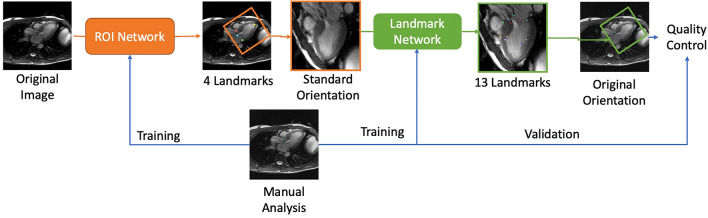
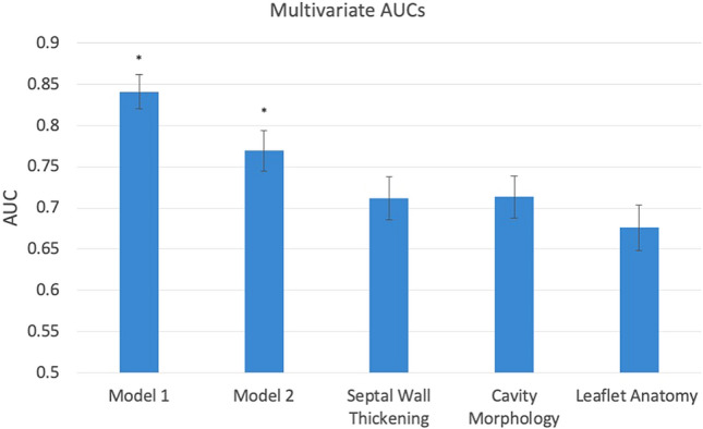
Left ventricular outflow tract obstruction (LVOTO) is common in hypertrophic cardiomyopathy (HCM), but relationships between anatomical metrics and obstruction are poorly understood. We aimed to develop machine learning methods to evaluate LVOTO in HCM patients and quantify relationships between anatomical metrics and obstruction. This retrospective analysis of 1905 participants of the HCM Registry quantified 11 anatomical metrics derived from 14 landmarks automatically detected on the three-chamber long axis cine CMR images. Linear and logistic regression was used to quantify strengths of relationships with the presence of LVOTO (defined by resting Doppler pressure drop of > 30 mmHg), using the area under the receiver operating characteristic (AUC). Intraclass correlation coefficients between the network predictions and three independent observers showed similar agreement to that between observers. The distance from anterior mitral valve leaflet tip to basal septum (AML-BS) was most highly correlated with Doppler pressure drop (R = 0.19, p < 10). Multivariate stepwise regression found the best predictive model included AML-BS, AML length to aortic valve diameter ratio, AML length to LV width ratio, and midventricular septal thickness metrics (AUC 0.84). Excluding AML-BS, metrics grouped according to septal hypertrophy, LV geometry, and AML anatomy each had similar associations with LVOTO (AUC 0.71, 0.71, 0.68 respectively, p = ns), significantly less than their combination (AUC 0.77, p < 0.05 for each). Anatomical metrics derived from a standard three-chamber CMR cine acquisition can be used to highlight risk of LVOTO, and suggest further investigation if necessary. A combination of geometric factors is required to provide the best risk prediction.

摘要

左心室流出道梗阻(LVOTO)在肥厚型心肌病(HCM)中很常见,但解剖学指标与梗阻之间的关系尚不清楚。我们旨在开发机器学习方法来评估 HCM 患者的 LVOTO 并量化解剖学指标与梗阻之间的关系。这项对 HCM 登记处 1905 名参与者的回顾性分析,使用来自自动检测的 14 个标记的三个心腔长轴电影 CMR 图像,量化了 11 个解剖学指标。线性和逻辑回归用于量化存在 LVOTO(定义为休息时多普勒压力降> 30mmHg)与解剖学指标之间的关系强度,使用接收者操作特征曲线下的面积(AUC)。网络预测与三个独立观察者之间的组内相关系数表明,与观察者之间的一致性相似。前二尖瓣叶尖端到基底部间隔(AML-BS)与多普勒压力降的相关性最高(R = 0.19,p < 10)。多变量逐步回归发现,最佳预测模型包括 AML-BS、AML 长度与主动脉瓣直径比、AML 长度与 LV 宽度比以及中隔厚度指标(AUC 0.84)。排除 AML-BS 后,根据间隔肥厚、LV 几何形状和 AML 解剖分组的指标与 LVOTO 具有相似的相关性(AUC 分别为 0.71、0.71 和 0.68,p = ns),显著低于其组合(AUC 0.77,p < 0.05)。来自标准三腔 CMR 电影采集的解剖学指标可用于突出 LVOTO 的风险,如果有必要,还可以进一步调查。需要组合几何因素以提供最佳风险预测。

https://cdn.ncbi.nlm.nih.gov/pmc/blobs/6955/9708771/b1ca6e3b6a30/10554_2022_2724_Fig4_HTML.jpg
https://cdn.ncbi.nlm.nih.gov/pmc/blobs/6955/9708771/92ba88ff5ea5/10554_2022_2724_Fig1_HTML.jpg
https://cdn.ncbi.nlm.nih.gov/pmc/blobs/6955/9708771/42dc974c8e72/10554_2022_2724_Fig2_HTML.jpg
https://cdn.ncbi.nlm.nih.gov/pmc/blobs/6955/9708771/bc5dc666d06e/10554_2022_2724_Fig3_HTML.jpg
https://cdn.ncbi.nlm.nih.gov/pmc/blobs/6955/9708771/b1ca6e3b6a30/10554_2022_2724_Fig4_HTML.jpg

相似文献

1
Machine learning evaluation of LV outflow obstruction in hypertrophic cardiomyopathy using three-chamber cardiovascular magnetic resonance.使用三房心心血管磁共振评估肥厚型心肌病的左心室流出道梗阻的机器学习方法。
Int J Cardiovasc Imaging. 2022 Dec;38(12):2695-2705. doi: 10.1007/s10554-022-02724-7. Epub 2022 Oct 6.
2
Contribution of mitral valve leaflet length and septal wall thickness to outflow tract obstruction in patients with hypertrophic cardiomyopathy.肥厚型心肌病患者二尖瓣叶长度和室间隔厚度对流出道梗阻的影响
Int J Cardiovasc Imaging. 2017 Aug;33(8):1201-1211. doi: 10.1007/s10554-017-1103-5. Epub 2017 Apr 8.
3
Evaluation of potential links between phenotypic features and genetic variants in left ventricular outflow tract obstruction in hypertrophic cardiomyopathy using cardiovascular magnetic resonance imaging.运用心血管磁共振成像评估肥厚型心肌病左心室流出道梗阻的表型特征与基因变异之间的潜在关联。
Int J Cardiovasc Imaging. 2024 Nov;40(11):2417-2428. doi: 10.1007/s10554-024-03250-4. Epub 2024 Sep 30.
4
Long mitral valve leaflets determine left ventricular outflow tract obstruction during exercise in hypertrophic cardiomyopathy.在肥厚型心肌病中,二尖瓣叶冗长决定运动时左心室流出道梗阻情况。
Int J Cardiol. 2016 Jun 1;212:47-53. doi: 10.1016/j.ijcard.2016.03.041. Epub 2016 Mar 16.
5
Spatial relationship between mitral valve and ventricular septum assessed by resting echocardiography to diagnose left ventricular outflow tract obstruction in hypertrophic cardiomyopathy.应用静息超声心动图评估二尖瓣与室间隔的空间关系诊断肥厚型心肌病患者左心室流出道梗阻。
Eur Heart J Cardiovasc Imaging. 2023 May 31;24(6):710-718. doi: 10.1093/ehjci/jead036.
6
Relationship of basal-septal fibrosis with LV outflow tract obstruction in hypertrophic cardiomyopathy: insights from cardiac magnetic resonance analysis.肥厚型心肌病中基底间隔纤维化与左心室流出道梗阻的关系:心脏磁共振分析的见解
Int J Cardiovasc Imaging. 2016 Apr;32(4):613-20. doi: 10.1007/s10554-015-0806-8. Epub 2015 Nov 20.
7
Geometric predictors of left ventricular outflow tract obstruction in patients with hypertrophic cardiomyopathy: a 3D computed tomography analysis.肥厚型心肌病患者左心室流出道梗阻的几何预测因素:3D 计算机断层扫描分析。
Eur Heart J Cardiovasc Imaging. 2018 Oct 1;19(10):1149-1156. doi: 10.1093/ehjci/jex234.
8
Left Ventricular Outflow Tract Obstruction in Hypertrophic Cardiomyopathy Patients Without Severe Septal Hypertrophy: Implications of Mitral Valve and Papillary Muscle Abnormalities Assessed Using Cardiac Magnetic Resonance and Echocardiography.无严重室间隔肥厚的肥厚型心肌病患者的左心室流出道梗阻:利用心脏磁共振成像和超声心动图评估二尖瓣和乳头肌异常的意义
Circ Cardiovasc Imaging. 2015 Jul;8(7):e003132. doi: 10.1161/CIRCIMAGING.115.003132.
9
Quantification of mitral regurgitation in patients with hypertrophic cardiomyopathy using aortic and pulmonary flow data: impacts of left ventricular outflow tract obstruction and different left ventricular segmentation methods.应用主动脉和肺血流数据对肥厚型心肌病患者二尖瓣反流进行定量评估:左心室流出道梗阻和不同左心室分段方法的影响。
J Cardiovasc Magn Reson. 2017 Dec 21;19(1):105. doi: 10.1186/s12968-017-0417-8.
10
Relation between left ventricular outflow tract obstruction and left ventricular shape in patients with hypertrophic cardiomyopathy: a cardiac magnetic resonance imaging study.肥厚型心肌病患者左心室流出道梗阻与左心室形态的关系:一项心脏磁共振成像研究。
Arch Cardiovasc Dis. 2013 Aug-Sep;106(8-9):440-7. doi: 10.1016/j.acvd.2013.05.002. Epub 2013 Jul 29.

引用本文的文献

1
Artificial Intelligence in Cardiovascular Imaging: Current Applications and New Horizons.心血管成像中的人工智能:当前应用与新视野
J Cardiovasc Echogr. 2025 Apr-Jun;35(2):97-107. doi: 10.4103/jcecho.jcecho_62_25. Epub 2025 Jul 30.
2
Exploring the Current Status of Risk Stratification in Hypertrophic Cardiomyopathy: From Risk Models to Promising Techniques.探索肥厚型心肌病风险分层的现状:从风险模型到有前景的技术。
J Cardiovasc Dev Dis. 2025 Mar 14;12(3):101. doi: 10.3390/jcdd12030101.
3
Spatial relationship between mitral valve and ventricular septum assessed by resting echocardiography to diagnose left ventricular outflow tract obstruction in hypertrophic cardiomyopathy.

本文引用的文献

1
Diagnosis and Evaluation of Hypertrophic Cardiomyopathy: JACC State-of-the-Art Review.肥厚型心肌病的诊断与评估:美国心脏病学会心血管造影与介入学会最新临床综述
J Am Coll Cardiol. 2022 Feb 1;79(4):372-389. doi: 10.1016/j.jacc.2021.12.002.
2
Evaluation of aortic stenosis: From Bernoulli and Doppler to Navier-Stokes.主动脉瓣狭窄的评估:从伯努利和多普勒到纳维-斯托克斯。
Trends Cardiovasc Med. 2023 Jan;33(1):32-43. doi: 10.1016/j.tcm.2021.12.003. Epub 2021 Dec 15.
3
Distinct Subgroups in Hypertrophic Cardiomyopathy in the NHLBI HCM Registry.肥厚型心肌病 NHLBI 登记处的不同亚组。
应用静息超声心动图评估二尖瓣与室间隔的空间关系诊断肥厚型心肌病患者左心室流出道梗阻。
Eur Heart J Cardiovasc Imaging. 2023 May 31;24(6):710-718. doi: 10.1093/ehjci/jead036.
4
Left ventricular anatomy in obstructive hypertrophic cardiomyopathy: beyond basal septal hypertrophy.梗阻性肥厚型心肌病的左心室解剖结构:不仅仅是基底室间隔肥厚。
Eur Heart J Cardiovasc Imaging. 2023 May 31;24(6):807-818. doi: 10.1093/ehjci/jeac233.
5
Editor's choice to the December 2022 issue : Machine learning evaluation of LV outflow obstruction in hypertrophic cardiomyopathy using threechamber cardiovascular magnetic resonance.
Int J Cardiovasc Imaging. 2022 Dec;38(12):2511-2512. doi: 10.1007/s10554-022-02758-x.
J Am Coll Cardiol. 2019 Nov 12;74(19):2333-2345. doi: 10.1016/j.jacc.2019.08.1057.
4
Clinical Course and Management of Hypertrophic Cardiomyopathy.肥厚型心肌病的临床病程与管理
N Engl J Med. 2018 Aug 16;379(7):655-668. doi: 10.1056/NEJMra1710575.
5
Morphological Determinants of Obstructive Hypertrophic Cardiomyopathy Obtained Using Echocardiography.使用超声心动图获得的梗阻性肥厚型心肌病的形态学决定因素。
Int Heart J. 2018 Mar 30;59(2):339-346. doi: 10.1536/ihj.17-072. Epub 2018 Feb 23.
6
Awareness of 'Systolic Anterior Motion' in Different Conditions.不同情况下对“收缩期前向运动”的认识
Clin Med Insights Cardiol. 2018 Jan 10;12:1179546817751921. doi: 10.1177/1179546817751921. eCollection 2018.
7
Discrepant Measurements of Maximal Left Ventricular Wall Thickness Between Cardiac Magnetic Resonance Imaging and Echocardiography in Patients With Hypertrophic Cardiomyopathy.肥厚型心肌病患者心脏磁共振成像与超声心动图测量左心室最大壁厚的差异
Circ Cardiovasc Imaging. 2017 Aug;10(8). doi: 10.1161/CIRCIMAGING.117.006309.
8
Comparison of Ventricular Septal Measurements in Hypertrophic Cardiomyopathy Patients Who Underwent Surgical Myectomy Using Multimodality Imaging and Implications for Diagnosis and Management.肥厚型心肌病患者行外科心肌切除术时室间隔测量的多模态影像学比较及其对诊断和管理的意义
Am J Cardiol. 2017 May 15;119(10):1656-1662. doi: 10.1016/j.amjcard.2017.02.009. Epub 2017 Mar 1.
9
Beyond Bernoulli: Improving the Accuracy and Precision of Noninvasive Estimation of Peak Pressure Drops.超越伯努利原理:提高无创估计峰值压力降的准确性和精度。
Circ Cardiovasc Imaging. 2017 Jan;10(1):e005207. doi: 10.1161/CIRCIMAGING.116.005207.
10
Comparison of Maximal Wall Thickness in Hypertrophic Cardiomyopathy Differs Between Magnetic Resonance Imaging and Transthoracic Echocardiography.肥厚型心肌病最大室壁厚度在磁共振成像和经胸超声心动图检查中的比较存在差异。
Am J Cardiol. 2017 Feb 15;119(4):643-650. doi: 10.1016/j.amjcard.2016.11.010. Epub 2016 Nov 16.