Pas Maciej, Jogo Atsushi, Yamamoto Akira, Nishida Norifumi, Jogo Eri, Kageyama Ken, Sohgawa Etsuji, Miki Yukio
Department of Diagnostic and Interventional Radiology, Graduate School of Medicine, Osaka Metropolitan University, 1-4-3 Asahi-machi, Abeno-ku, Osaka 545-8585, Japan.
Department of Radiology, Saiseikai Nakatsu Hospital, 2-10-39 Shibata, Kita-ward, Osaka City, Osaka, 530-0012, Japan.
Radiol Case Rep. 2022 Sep 30;17(12):4679-4684. doi: 10.1016/j.radcr.2022.08.096. eCollection 2022 Dec.
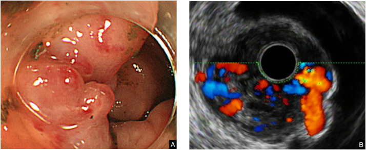
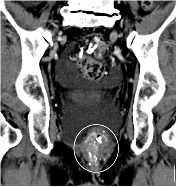
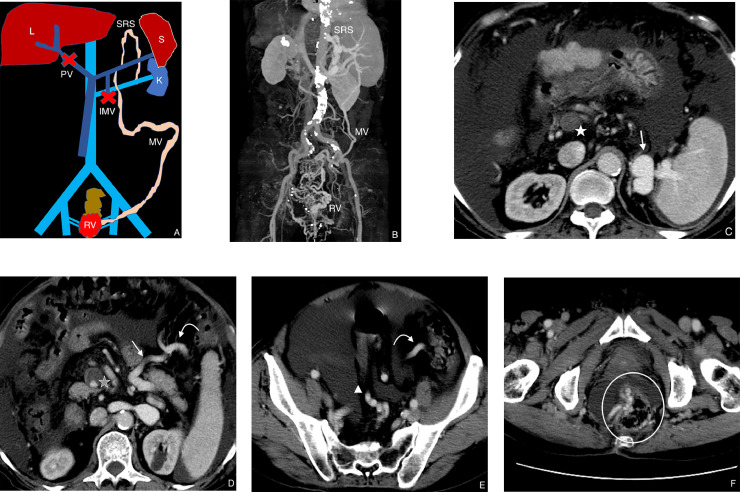
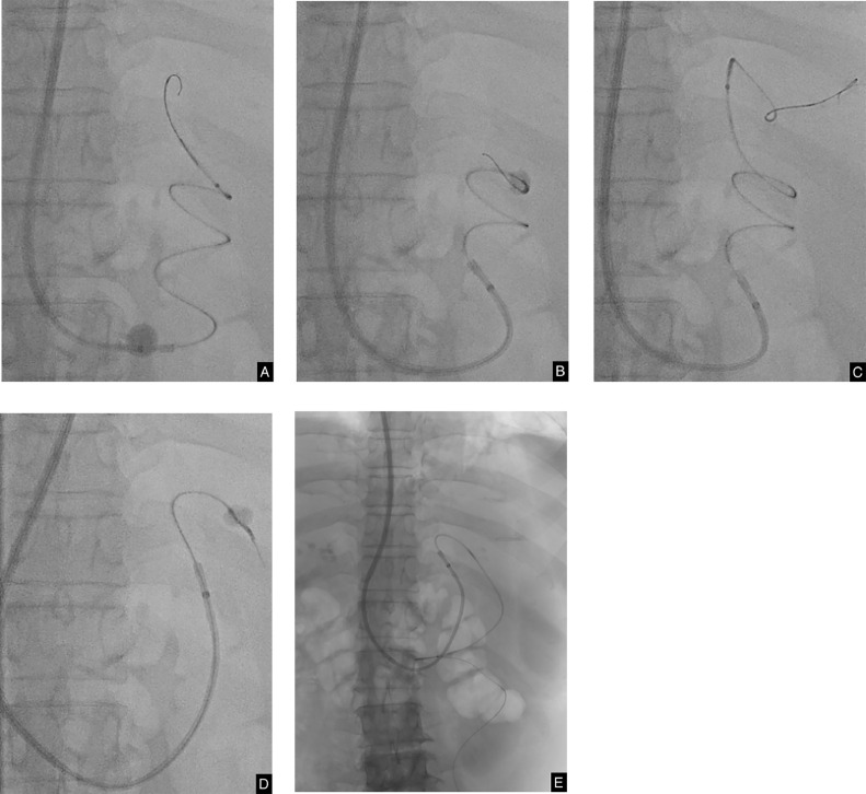
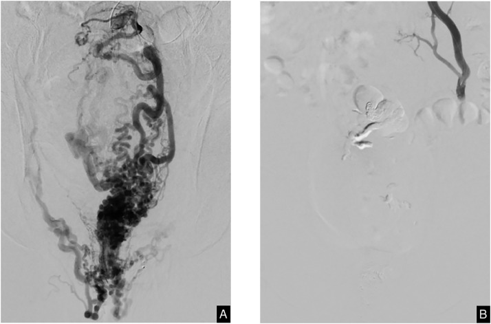
A 74-year-old patient presented with hematochezia and a history of liver cirrhosis with repeated bleeding from esophageal and rectal varices. Endoscopic examination revealed multiple rectal varices with positive red color signs. Ascites, severe portosystemic thrombosis and a splenorenal shunt were diagnosed on a contrast-enhanced dynamic computed tomography examination. From a transjugular approach, we circumvented thrombosed regions by maneuvering double balloon catheters through the shunt and dilated left colic marginal vein. We managed to successfully obliterate the varices.
一名74岁患者出现便血,有肝硬化病史,曾反复出现食管和直肠静脉曲张出血。内镜检查发现多处直肠静脉曲张,红色征阳性。在增强动态计算机断层扫描检查中诊断出腹水、严重的门体系统血栓形成和脾肾分流。我们采用经颈静脉途径,通过分流处操纵双气囊导管避开血栓形成区域,并扩张左结肠边缘静脉。我们成功地消除了静脉曲张。