Zhang Zixuan, Fang Qiong, Zhang Yu, Zhu Youzhi, Zhang Wei, Zhu Youyou, Deng Xuefei
Department of Clinical Medicine, West Anhui Health Vocational College, Lu'an, China.
Department of Anatomy, Anhui Medical University, Hefei, China.
Front Cardiovasc Med. 2022 Sep 21;9:1013610. doi: 10.3389/fcvm.2022.1013610. eCollection 2022.
Arterial spasm is proved to be an inducer of cerebral ischemia and cerebral infarction, while when a venous spasm occurs, cerebral edema is seen to be caused by a disturbance in cerebral blood flow. However, it is unclear and unproven whether venous spasm occurs after subarachnoid hemorrhage (SAH). To provide the theoretical basis for treating cerebral vasospasm after SAH, magnetic resonance imaging (MRI) was employed to observe the changes in the diameter of deep cerebral veins in rabbits after SAH.
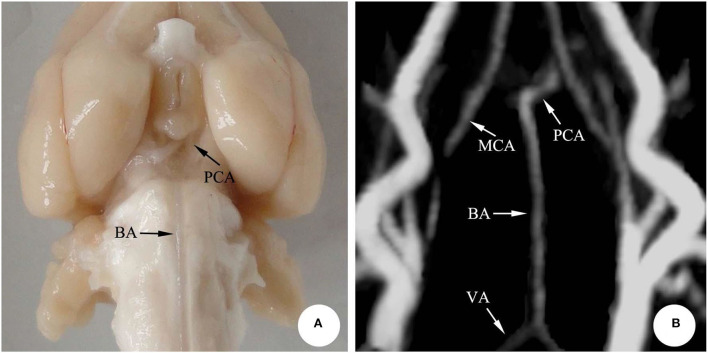
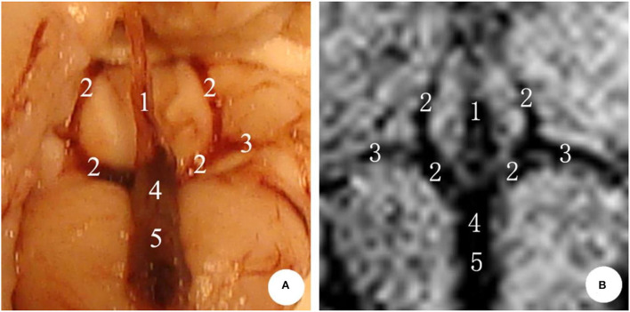
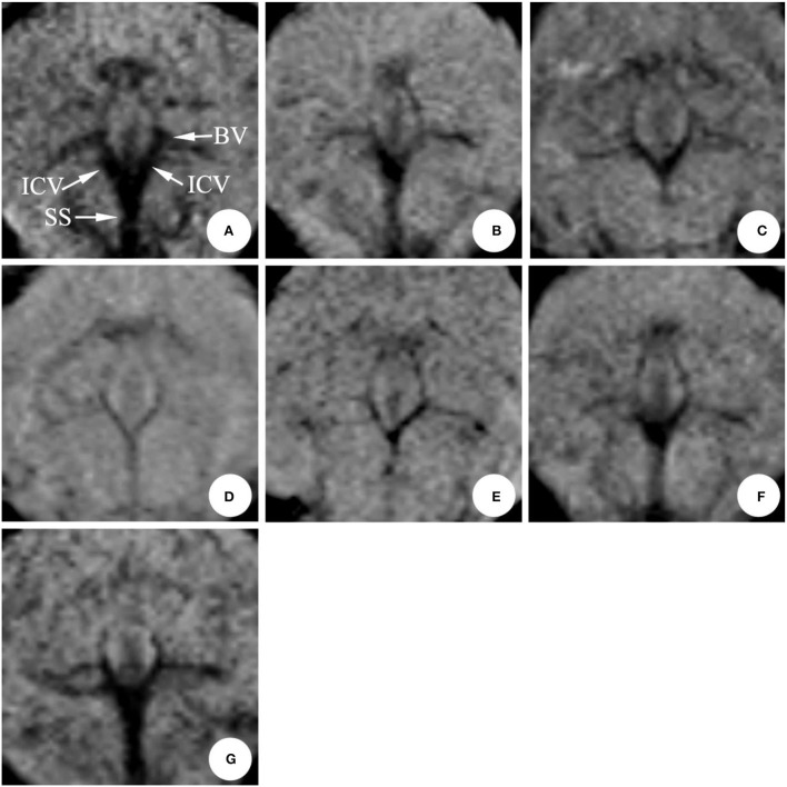
Fourteen New Zealand rabbits were randomly divided into the SAH group ( = 10) and the normal saline group (NS group, = 4). Specifically, the SAH models were established by the ultrasound-guided double injections of blood into cisterna magna. Moreover, the MRI was performed to observe the changes in the diameter of deep cerebral veins (internal cerebral vein, basilar vein, and great cerebral vein) and basilar artery before modeling (0 d) and 1, 3, 5, 7, 9, and 11 d after modeling.
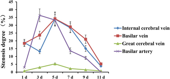
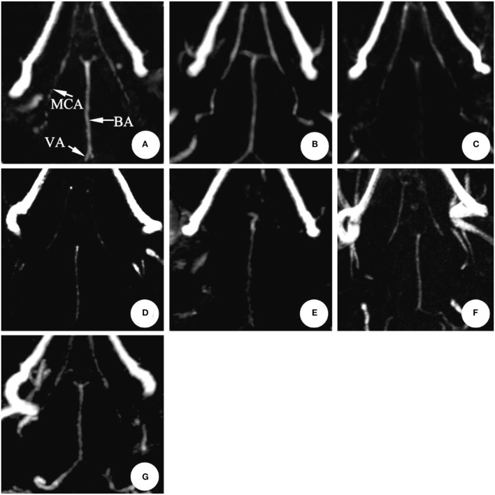
In the SAH group, the diameter of the basilar artery showed no evident change on the 1st d. However, it became narrower obviously on the 3rd d and 5th d, and the stenosis degree was more than 30%. The diameter gradually relieved from 7th to 9th d, and finally returned to normal on the 11th d. Moreover, the diameter of the internal cerebral vein significantly narrowed on the 1st d, the stenosis degree of which was 19%; the stenosis then relieved slightly on the 3rd d (13%), reached the peak (34%) on the 5th d, and gradually relieved from 7th d to 11th d. Moreover, the stenosis degree of the basilar vein was 18% on the 1st d, 24% on the 3rd d, and reached the peak (34%) on the 5th d.
After SAH in rabbits, the cerebral vasospasm was seen to occur in the basilar artery, and likewise, spasmodic changes took place in the deep cerebral vein. Furthermore, the time regularity of spasmodic changes between the cerebral vein and basilar artery was of significant difference, indicating that the venous vasospasm resulted in active contraction.
动脉痉挛被证实是脑缺血和脑梗死的诱因,而静脉痉挛发生时,脑水肿被认为是由脑血流紊乱引起的。然而,蛛网膜下腔出血(SAH)后是否会发生静脉痉挛尚不清楚且未经证实。为了为SAH后脑血管痉挛的治疗提供理论依据,采用磁共振成像(MRI)观察SAH后家兔大脑深静脉直径的变化。
将14只新西兰兔随机分为SAH组(n = 10)和生理盐水组(NS组,n = 4)。具体而言,通过超声引导向枕大池双次注入血液建立SAH模型。此外,在建模前(0 d)以及建模后1、3、5、7、9和11 d进行MRI,观察大脑深静脉(大脑内静脉、基底静脉和大脑大静脉)和基底动脉直径的变化。
在SAH组中,基底动脉直径在第1天无明显变化。然而,在第3天和第5天明显变窄,狭窄程度超过30%。直径在第7天至第9天逐渐缓解,最终在第11天恢复正常。此外,大脑内静脉直径在第1天显著变窄,狭窄程度为19%;然后在第3天略有缓解(13%),在第5天达到峰值(34%),并从第7天至第11天逐渐缓解。此外,基底静脉狭窄程度在第1天为18%,第3天为24%,并在第5天达到峰值(34%)。
家兔SAH后,基底动脉出现脑血管痉挛,同样,大脑深静脉也发生痉挛性变化。此外,脑静脉和基底动脉痉挛性变化的时间规律存在显著差异,表明静脉血管痉挛导致了主动收缩。