Merkel Helena, Lindner Dirk, Gaber Khaled, Ziganshyna Svitlana, Jentzsch Jennifer, Mucha Simone, Gerhards Thilo, Sari Sabine, Stock Annika, Vothel Felicitas, Falter Lea, Quäschling Ulf, Hoffmann Karl-Titus, Meixensberger Jürgen, Halama Dirk, Richter Cindy
Department of Neuroradiology, Leipzig University Hospital, Liebigstraße 20, 04103 Leipzig, Germany.
Department of Neurosurgery, Leipzig University Hospital, Liebigstraße 20, 04103 Leipzig, Germany.
J Clin Med. 2022 Apr 3;11(7):2011. doi: 10.3390/jcm11072011.
During the last decade, cerebral vasospasm after aneurysmal subarachnoid hemorrhage (SAH) was a current research focus without a standardized classification in digital subtraction angiography (DSA). This study was performed to investigate a device-independent visual cerebral vasospasm classification for endovascular treatment.
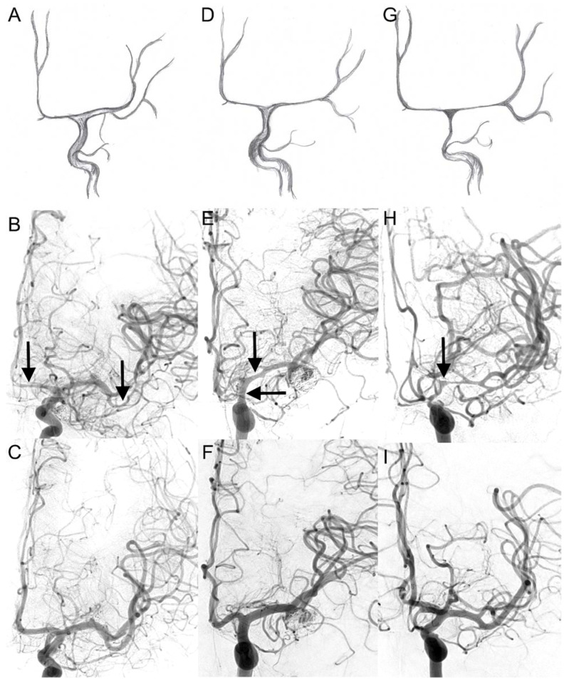
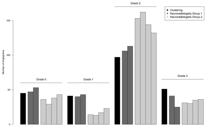
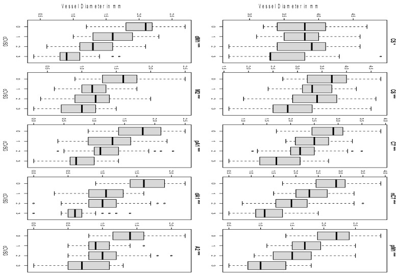
The analyses are DSA based rather than multimodal. Ten defined points of intracranial arteries were measured in 45 patients suffering from cerebral vasospasm after SAH at three time points (hospitalization, before spasmolysis, control after six months). Mathematical clustering of vessel diameters was performed to generate four objective grades for comparison. Six interventional neuroradiologists in two groups scored 237 DSAs after a new visual classification (grade 0-3) developed on a segmental pattern of vessel contraction. For the second group, a threshold-based criterion was amended.
The raters had a reproducibility of 68.4% in the first group and 75.2% in the second group. The complementary threshold-based criterion increased the reproducibility by about 6.8%, while the rating deviated more from the mathematical clustering in all grades.
The proposed visual classification scheme of cerebral vasospasm is suitable as a standard grading procedure for endovascular treatment. There is no advantage of a threshold-based criterion that compensates for the effort involved. Automated vessel analysis is superior to compare inter-group results in research settings.
在过去十年中,动脉瘤性蛛网膜下腔出血(SAH)后的脑血管痉挛是当前的研究热点,在数字减影血管造影(DSA)中尚无标准化分类。本研究旨在探讨一种独立于设备的视觉脑血管痉挛分类方法,用于血管内治疗。
分析基于DSA而非多模态。在45例SAH后发生脑血管痉挛的患者中,于三个时间点(住院时、血管痉挛解除前、六个月后复查)测量颅内动脉的10个定义点。对血管直径进行数学聚类以生成四个客观等级进行比较。两组的六名介入神经放射科医生根据基于血管收缩节段模式制定的新视觉分类(0 - 3级)对237份DSA进行评分。对于第二组,修改了基于阈值的标准。
第一组评估者的再现性为68.4%,第二组为75.2%。基于阈值的补充标准使再现性提高了约6.8%,而在所有等级中评分与数学聚类的偏差更大。
所提出的脑血管痉挛视觉分类方案适合作为血管内治疗的标准分级程序。基于阈值的标准并无优势,且需付出额外努力。在研究环境中,自动血管分析在比较组间结果方面更具优势。