Farid Mariam F, Abouelela Yara S, Yasin Noha A E, Mousa Mohamed R, Ibrahim Marwa A, Prince Abdelbary, Rizk Hamdy
Anatomy and Embryology Department, Faculty of Veterinary Medicine, Cairo University, Giza, 12211, Egypt.
Cytology and Histology Department, Faculty of Veterinary Medicine, Cairo University, Giza, Egypt.
Inflamm Regen. 2022 Oct 14;42(1):45. doi: 10.1186/s41232-022-00230-w.
Multiple sclerosis (MS) is a progressive autoimmune demyelinating disease of the central nervous system. To date, there is no effective therapy for it. Our study aimed to determine the potential role of platelet-rich plasma (PRP) in the treatment of MS in cats.
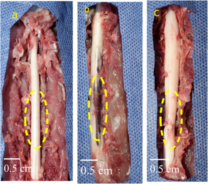
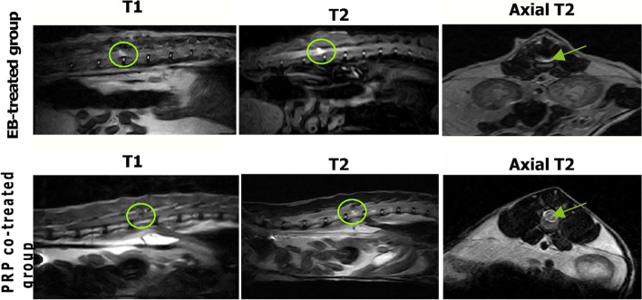
The current study was conducted on 15 adult Persian cats that were divided into three groups: control negative, control positive (ethidium bromide (EB)-treated group), and PRP co-treated group (EB-treated group intrathecally injected with PRP on day 14 post-spinal cord injury). PRP was obtained by centrifuging blood on anticoagulant citrate dextrose and activating it with red and green laser diodes. The Basso-Beattie-Bresnahan (BBB) scores were used to assess the motor function recovery on days 1, 3, 7, 14, 20, and 28 following 14 days from EB injection. Moreover, magnetic resonance imaging (MRI) analysis, histopathological investigations, transmission electron microscopy (TEM) studies, and immunohistochemical analysis were conducted, and the gene expressions of nerve growth factors (NGFs), brain-derived neurotrophic factors (BDNF), and stromal cell-derived factors (SDF) were evaluated.
Our results indicated that PRP had a significant ameliorative effect on the motor function of the hindlimbs as early as day 20 and so on. MRI revealed that the size and intensity of the lesion were significantly reduced in the PRP co-treated group. The histopathological and TEM investigations demonstrated that the PRP co-treated group had a significant improvement in the structure and organization of the white matter, as well as a high remyelination capacity. Furthermore, a significant increase in myelin basic protein and Olig2 immunoreactivity as well as a reduction in Bax and glial fibrillar acidic protein immune markers was observed. NGFs were found to be upregulated by gene expression.
As a result, we concluded that the intrathecal injection of PRP was an effective, safe, and promising method for the treatment of MS.
多发性硬化症(MS)是一种中枢神经系统的进行性自身免疫性脱髓鞘疾病。迄今为止,尚无有效的治疗方法。我们的研究旨在确定富血小板血浆(PRP)在猫MS治疗中的潜在作用。
本研究对15只成年波斯猫进行,分为三组:阴性对照组、阳性对照组(溴化乙锭(EB)处理组)和PRP联合处理组(脊髓损伤后第14天对EB处理组进行鞘内注射PRP)。通过在枸橼酸葡萄糖抗凝剂上离心血液并使用红色和绿色激光二极管激活来获得PRP。在EB注射14天后的第1、3、7、14、20和28天,使用Basso-Beattie-Bresnahan(BBB)评分评估运动功能恢复情况。此外,进行了磁共振成像(MRI)分析、组织病理学研究、透射电子显微镜(TEM)研究和免疫组织化学分析,并评估了神经生长因子(NGF)、脑源性神经营养因子(BDNF)和基质细胞衍生因子(SDF)的基因表达。
我们的结果表明,PRP早在第20天等就对后肢运动功能有显著改善作用。MRI显示,PRP联合处理组病变的大小和强度显著降低。组织病理学和TEM研究表明,PRP联合处理组在白质结构和组织方面有显著改善,且髓鞘再生能力较高。此外,观察到髓鞘碱性蛋白和Olig2免疫反应性显著增加,以及Bax和胶质纤维酸性蛋白免疫标志物减少。发现NGF通过基因表达上调。
因此,我们得出结论,鞘内注射PRP是一种治疗MS的有效、安全且有前景的方法。