Bütof Rebecca, Hönscheid Pia, Aktar Rozina, Sperling Christian, Tillner Falk, Rassamegevanon Treewut, Dietrich Antje, Meinhardt Matthias, Aust Daniela, Krause Mechthild, Troost Esther G C
OncoRay-National Center for Radiation Research in Oncology, Faculty of Medicine and University Hospital Carl Gustav Carus, Technische Universität Dresden, Helmholtz-Zentrum Dresden-Rossendorf, 01307 Dresden, Germany.
Department of Radiotherapy and Radiation Oncology, Faculty of Medicine and University Hospital Carl Gustav Carus, Technische Universität Dresden, 01307 Dresden, Germany.
Cancers (Basel). 2022 Sep 20;14(19):4559. doi: 10.3390/cancers14194559.
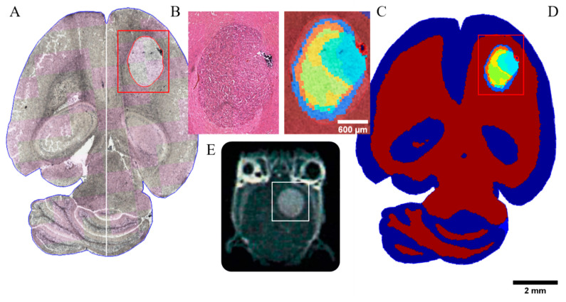
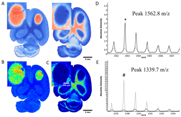
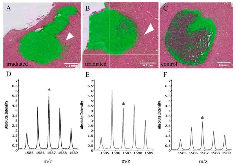
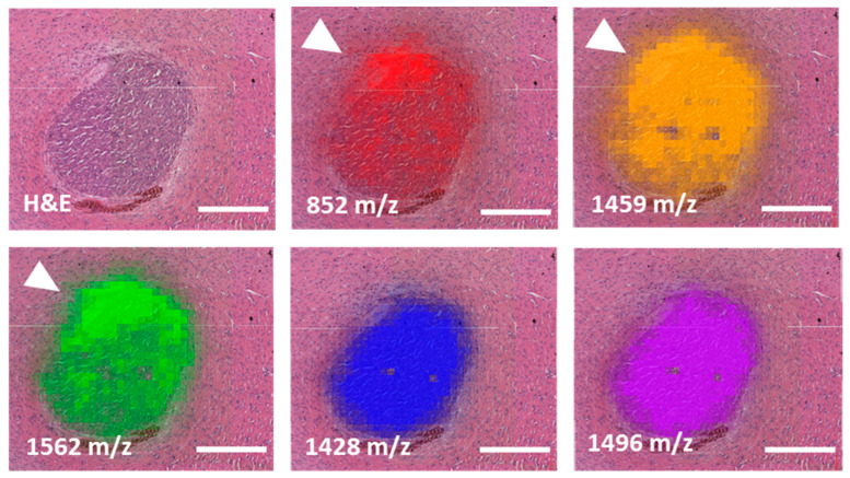
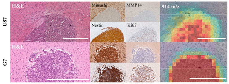
In times of high-precision radiotherapy, the accurate and precise definition of the primary tumor localization and its microscopic spread is of enormous importance. In glioblastoma, the microscopic tumor extension is uncertain and, therefore, population-based margins for Clinical Target Volume (CTV) definition are clinically used, which could either be too small-leading to increased risk of loco-regional recurrences-or too large, thus, enhancing the probability of normal tissue toxicity. Therefore, the aim of this project is to investigate an individualized definition of the CTV in preclinical glioblastoma models based on specific biological tumor characteristics. The microscopic tumor extensions of two different orthotopic brain tumor models (U87MG_mCherry; G7_mCherry) were evaluated before and during fractionated radiotherapy and correlated with corresponding histological data. Representative tumor slices were analyzed using Matrix-Assisted Laser Desorption/Ionization (MALDI) and stained for putative stem-like cell markers as well as invasion markers. The edges of the tumor are clearly shown by the MALDI segmentation via unsupervised clustering of mass spectra and are consistent with the histologically defined border in H&E staining in both models. MALDI component analysis identified specific peaks as potential markers for normal brain tissue (e.g., 1339 /), whereas other peaks demarcated the tumors very well (e.g., 1562 / for U87MG_mCherry) irrespective of treatment. MMP14 staining revealed only a few positive cells, mainly in the tumor border, which could reflect the invasive front in both models. The results of this study indicate that MALDI information correlates with microscopic tumor spread in glioblastoma models. Therefore, an individualized CTV definition based on biological tumor characteristics seems possible, whereby the visualization of tumor volume and protein heterogeneity can be potentially used to define radiotherapy-sensitive and resistant areas.
在高精度放射治疗时代,准确精确地定义原发性肿瘤定位及其微观扩散至关重要。在胶质母细胞瘤中,微观肿瘤扩展情况不确定,因此临床上使用基于人群的临床靶区(CTV)定义边界,这可能过小——导致局部区域复发风险增加——或者过大,从而增加正常组织毒性的可能性。因此,本项目的目的是在临床前胶质母细胞瘤模型中,基于特定的生物学肿瘤特征研究CTV的个体化定义。在分次放射治疗前和治疗期间,评估了两种不同的原位脑肿瘤模型(U87MG_mCherry;G7_mCherry)的微观肿瘤扩展情况,并将其与相应的组织学数据相关联。使用基质辅助激光解吸/电离(MALDI)分析代表性肿瘤切片,并对假定的干细胞样标志物以及侵袭标志物进行染色。通过对质谱进行无监督聚类的MALDI分割清晰显示了肿瘤边缘,且在两种模型中均与苏木精和伊红(H&E)染色中组织学定义的边界一致。MALDI成分分析确定了特定峰作为正常脑组织的潜在标志物(例如,1339 /),而其他峰能很好地界定肿瘤(例如,U87MG_mCherry的1562 /),与治疗无关。基质金属蛋白酶14(MMP14)染色仅显示少数阳性细胞,主要在肿瘤边界,这可能反映了两种模型中的侵袭前沿。本研究结果表明,MALDI信息与胶质母细胞瘤模型中的微观肿瘤扩散相关。因此,基于生物学肿瘤特征的个体化CTV定义似乎是可行的,由此肿瘤体积和蛋白质异质性的可视化可能用于定义放射治疗敏感和抵抗区域。