Maastricht Multimodal Molecular Imaging (M4I) Institute, Division of Imaging Mass Spectrometry, Maastricht University, Maastricht, The Netherlands.
Astrazeneca IMED-DSM, Cambridge, UK.
Mol Imaging Biol. 2018 Dec;20(6):888-901. doi: 10.1007/s11307-018-1267-y.
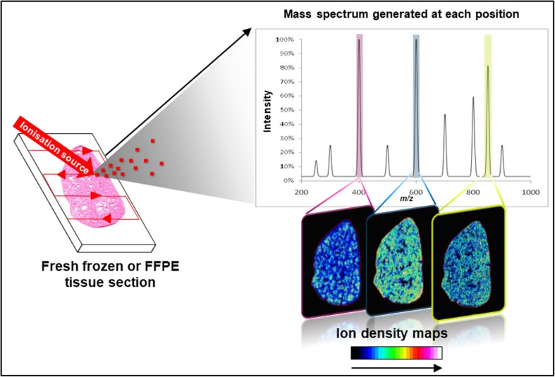
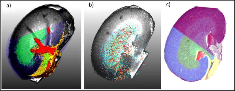
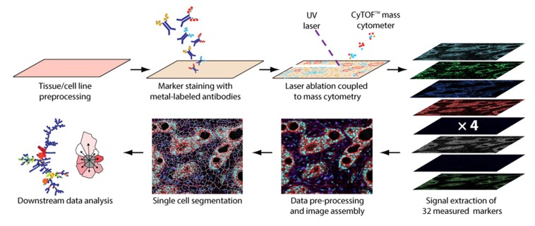
Over the last two decades, mass spectrometry imaging (MSI) has been increasingly employed to investigate the spatial distribution of a wide variety of molecules in complex biological samples. MSI has demonstrated its potential in numerous applications from drug discovery, disease state evaluation through proteomic and/or metabolomic studies. Significant technological and methodological advancements have addressed natural limitations of the techniques, i.e., increased spatial resolution, increased detection sensitivity especially for large molecules, higher throughput analysis and data management. One of the next major evolutions of MSI is linked to the introduction of imaging mass cytometry (IMC). IMC is a multiplexed method for tissue phenotyping, imaging signalling pathway or cell marker assessment, at sub-cellular resolution (1 μm). It uses MSI to simultaneously detect and quantify up to 30 different antibodies within a tissue section. The combination of MSI with other molecular imaging techniques can also provide highly relevant complementary information to explore new scientific fields. Traditionally, classical histology (especially haematoxylin and eosin-stained sections) is overlaid with molecular profiles obtained by MSI. Thus, MSI-based molecular histology provides a snapshot of a tissue microenvironment and enables the correlation of drugs, metabolites, lipids, peptides or proteins with histological/pathological features or tissue substructures. Recently, many examples combining MSI with other imaging modalities such as fluorescence, confocal Raman spectroscopy and MRI have emerged. For instance, brain pathophysiology has been studied using both MRI and MSI, establishing correlations between in and ex vivo molecular imaging techniques. Endogenous metabolite and small peptide modulation were evaluated depending on disease state. Here, we review advanced 'hot topics' in MSI development and explore the combination of MSI with established molecular imaging techniques to improve our understanding of biological and pathophysiological processes.
在过去的二十年中,质谱成像(MSI)已越来越多地用于研究复杂生物样本中各种分子的空间分布。MSI 在从药物发现到疾病状态评估的各种应用中都显示出了其潜力,通过蛋白质组学和/或代谢组学研究。重大的技术和方法学进步解决了该技术的一些固有局限性,例如,提高了空间分辨率,提高了检测灵敏度(尤其是对大分子而言),提高了高通量分析和数据管理水平。MSI 的下一个重大发展之一与成像质谱流式细胞术(IMC)的引入有关。IMC 是一种用于组织表型分析的多重方法,可对细胞信号通路或细胞标志物进行亚细胞分辨率(1 μm)的成像。它使用 MSI 同时检测和定量组织切片中多达 30 种不同的抗体。MSI 与其他分子成像技术的结合还可以提供高度相关的互补信息,以探索新的科学领域。传统上,经典组织学(尤其是苏木精和伊红染色切片)与通过 MSI 获得的分子谱重叠。因此,基于 MSI 的分子组织学为组织微环境提供了一个快照,并能够将药物、代谢物、脂质、肽或蛋白质与组织学/病理学特征或组织亚结构相关联。最近,出现了许多将 MSI 与其他成像模式(例如荧光、共聚焦拉曼光谱和 MRI)相结合的示例。例如,使用 MRI 和 MSI 研究了脑病理生理学,建立了体内和体外分子成像技术之间的相关性。根据疾病状态评估了内源性代谢物和小肽的调节。在这里,我们回顾了 MSI 发展的高级“热门话题”,并探讨了将 MSI 与成熟的分子成像技术相结合,以增进我们对生物和病理生理过程的理解。