Shi's Center of Orthopedics and Traumatology, Shuguang Hospital Affiliated to Shanghai University of Traditional Chinese Medicine, Institute of Traumatology & Orthopedics, Shanghai Academy of Traditional Chinese Medicine, No. 528, Zhangheng Road, Shanghai, Pudong New Area, 201203, China.
Sichuan Province Orthopaedic Hospital, No. 132, West Section 1, First Ring Road, Chengdu, Sichuan Province, China.
J Orthop Surg Res. 2022 Oct 15;17(1):454. doi: 10.1186/s13018-022-03348-2.
We aimed to investigate the utility of Hounsfield units (HU) obtained from different regions of interest in opportunistic lumbar computed tomography (CT) to predict osteoporosis coupling with data of dual-energy X-ray absorptiometry (DXA).
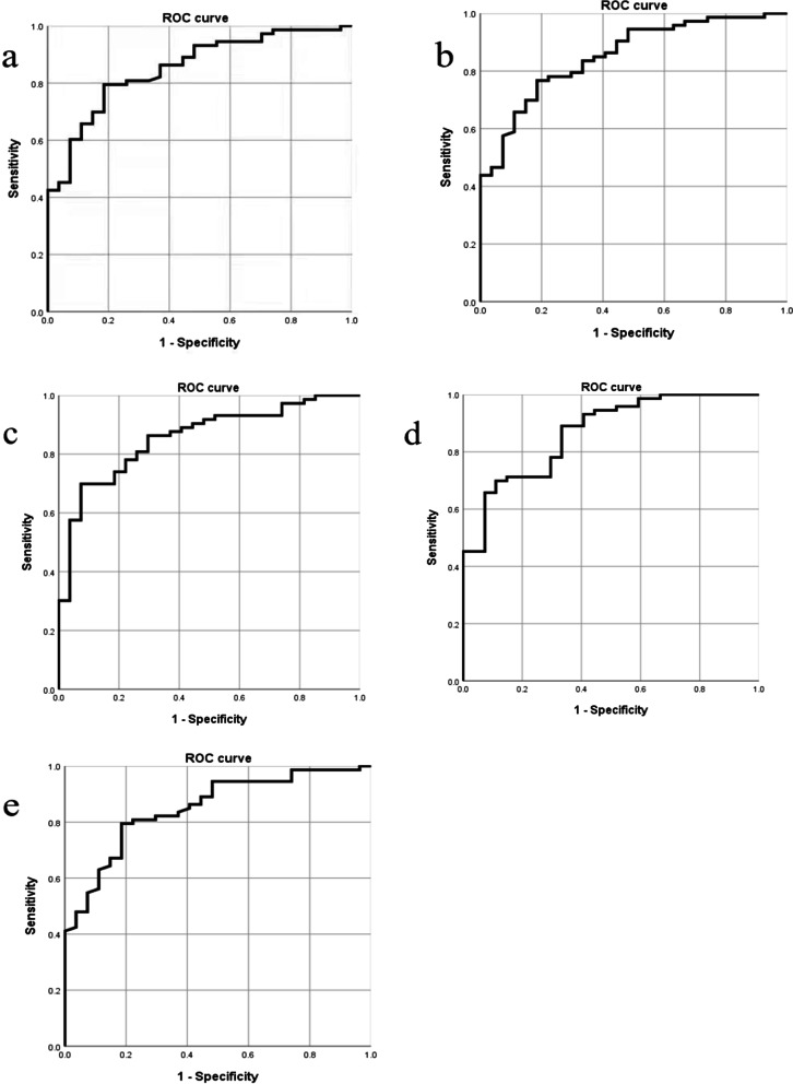
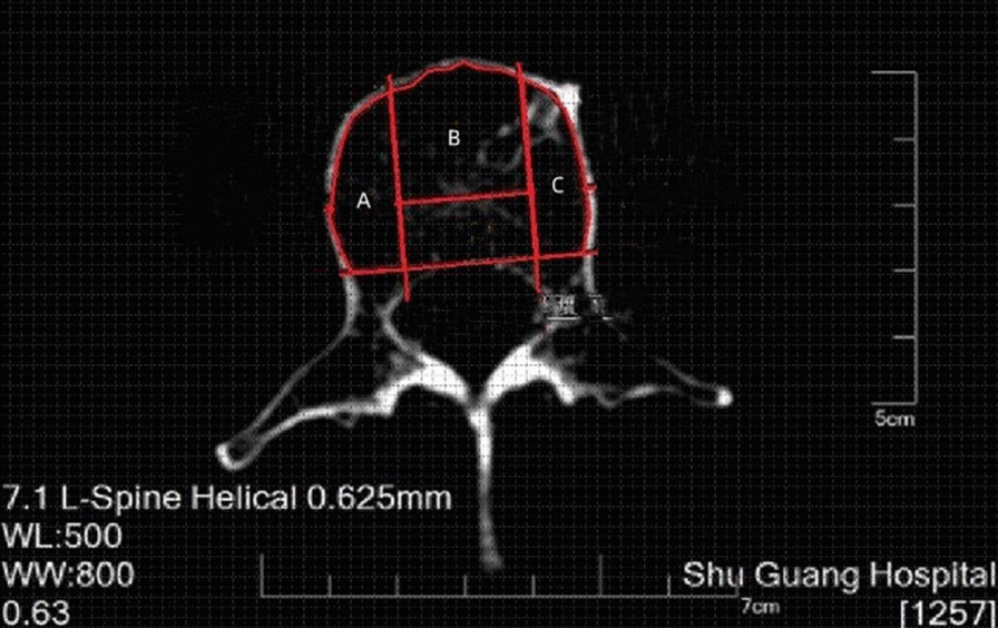
A total of 100 patients who attended a university hospital in Shanghai, China, and had undergone CT and DXA tests of the lumbar spine within 3 months were included in this retrospective review. Images were reviewed on axial sections, and regions of interest (ROI) markers were placed on the round, oval, anterior, left, and right of the L1-L4 vertebra to measure the HU. The mean values of CT HU were then compared to the bone mineral density (BMD) measured by DXA. Receiver operator characteristic curves were generated to determine the threshold for diagnosis and its sensitivity and specificity values.
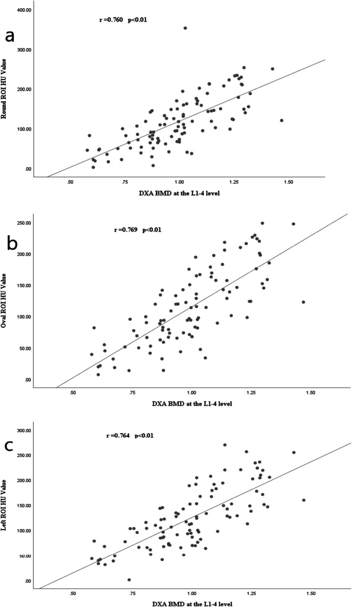
The differences in CT HU of different ROI based on DXA definitions of osteoporosis, osteopenia, and normal individuals were statistically significant (p < 0.01). The HU values of the different ROI correlated well with the BMD values (Spearman coefficient all > 0.75, p < 0.01). The threshold for diagnosing osteoporosis varies from 87 to 111 HU in different ROIs, and the threshold for excluding osteoporosis or osteopenia is 99-125 HU.
This is the first study on osteoporosis diagnosis of different ROI with routine CT lumbar scans. There is a strong correlation between CT HU of different ROI in the lumbar spine and BMD, and HU measurements can be used to predict osteoporosis.
我们旨在研究从机会性腰椎计算机断层扫描(CT)的不同感兴趣区获得的亨氏单位(HU)在多大程度上可用于预测骨质疏松症,并结合双能 X 射线吸收法(DXA)的数据。
本回顾性研究共纳入 100 例在上海某大学医院就诊并在 3 个月内同时接受过腰椎 CT 和 DXA 检查的患者。对轴向切片进行图像回顾,在 L1-L4 椎体的圆形、椭圆形、前、左、右侧放置感兴趣区(ROI)标记,以测量 HU。然后将 CT HU 的平均值与 DXA 测量的骨密度(BMD)进行比较。生成受试者工作特征曲线以确定诊断的阈值及其敏感性和特异性值。
根据 DXA 骨质疏松症、骨量减少和正常个体的定义,不同 ROI 的 CT HU 差异具有统计学意义(p<0.01)。不同 ROI 的 HU 值与 BMD 值相关性良好(Spearman 系数均>0.75,p<0.01)。不同 ROI 诊断骨质疏松症的阈值为 87-111 HU,排除骨质疏松症或骨量减少的阈值为 99-125 HU。
这是首次使用常规腰椎 CT 扫描对不同 ROI 的骨质疏松症进行诊断的研究。腰椎不同 ROI 的 CT HU 与 BMD 之间具有很强的相关性,HU 测量值可用于预测骨质疏松症。