Thau-Zuchman Orli, Pallier Patrick N, Savelkoul Paul J M, Kuipers Almar A M, Verkuyl J Martin, Michael-Titus Adina T
Centre for Neuroscience, Surgery and Trauma, Barts and The London School of Medicine and Dentistry, The Blizard Institute, Queen Mary University of London, London, United Kingdom.
Danone Nutricia Research, Utrecht, Netherlands.
Front Neurosci. 2022 Sep 29;16:926023. doi: 10.3389/fnins.2022.926023. eCollection 2022.
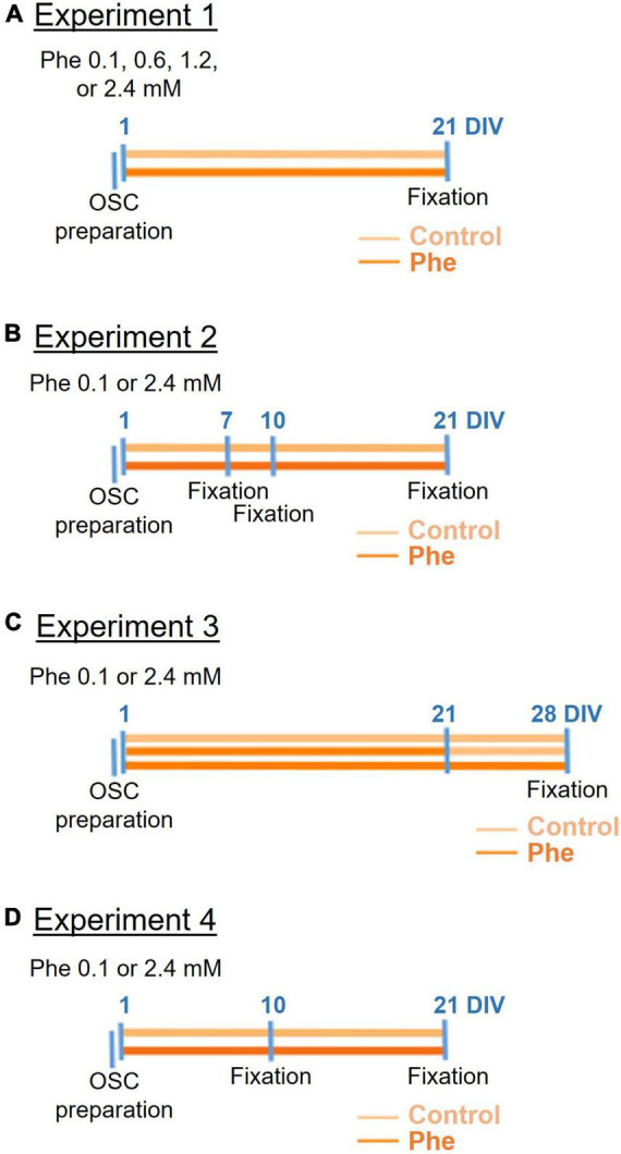
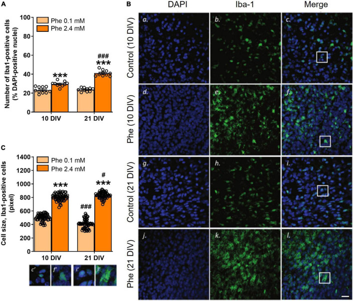
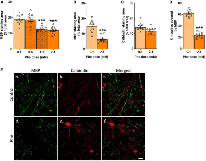
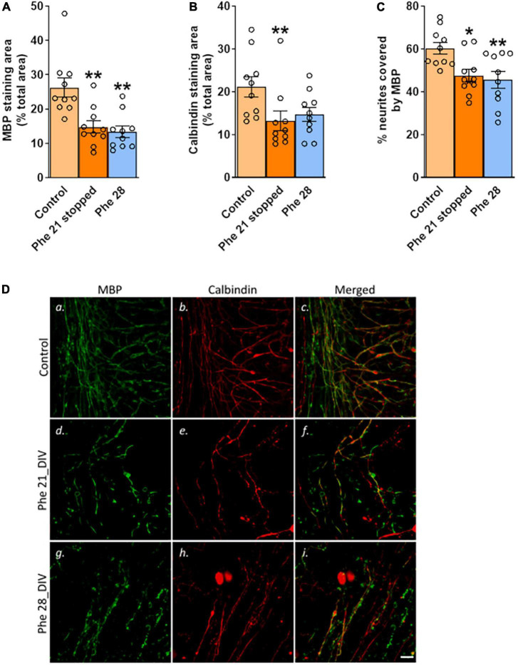
Phenylketonuria (PKU) is an inborn error of metabolism. Mutations in the enzyme phenylalanine hydroxylase (PAH)-encoding gene lead to a decreased metabolism of the amino acid phenylalanine (Phe). The deficiency in PAH increases Phe levels in blood and brain. Accumulation of Phe can lead to delayed development, psychiatric problems and cognitive impairment. White matter (WM) damage is a neuropathological hallmark of PKU and can be seen even in early detected and treated PKU patients. The mechanisms linking high Phe concentrations to WM abnormalities remain unclear. We tested the effects of high Phe concentrations on myelin in three models of increasing complexity: two simple cell culture models and one model that preserves local brain tissue architecture, a cerebellar organotypic slice culture prepared from postnatal day (P) 8 CD-1 mice. Various Phe concentrations (0.1-10 mM) and durations of exposure were tested. We found no toxic effect of high Phe in the cell culture models. On the contrary, the treatment promoted the maturation of oligodendrocytes, particularly at the highest, non-physiological Phe concentrations. Exposure of cerebellar organotypic slices to 2.4 mM Phe for 21 days (DIV), but not 7 or 10 DIV, resulted in a significant decrease in myelin basic protein (MBP), calbindin-stained neurites, and neurites co-stained with MBP. Following exposure to a toxic concentration of Phe, a switch to the control medium for 7 days did not lead to remyelination, while very active remyelination was seen in slices following demyelination with lysolecithin. An enhanced number of microglia, displaying an activated type morphology, was seen after exposure of the slices to 2.4 mM Phe for 10 or 21 DIV. The results suggest that prolonged exposure to high Phe concentrations can induce microglial activation preceding significant disruption of myelin.
苯丙酮尿症(PKU)是一种先天性代谢缺陷。编码苯丙氨酸羟化酶(PAH)的基因突变导致氨基酸苯丙氨酸(Phe)的代谢减少。PAH的缺乏会增加血液和大脑中的苯丙氨酸水平。苯丙氨酸的积累会导致发育迟缓、精神问题和认知障碍。白质(WM)损伤是PKU的神经病理学特征,即使在早期检测和治疗的PKU患者中也可见到。高苯丙氨酸浓度与白质异常之间的联系机制尚不清楚。我们在三个复杂性不断增加的模型中测试了高苯丙氨酸浓度对髓鞘的影响:两个简单的细胞培养模型和一个保留局部脑组织结构的模型,即从出生后第8天(P)的CD-1小鼠制备的小脑器官型切片培养物。测试了各种苯丙氨酸浓度(0.1-10 mM)和暴露持续时间。我们发现在细胞培养模型中高苯丙氨酸没有毒性作用。相反,这种处理促进了少突胶质细胞的成熟,特别是在最高的非生理苯丙氨酸浓度下。将小脑器官型切片暴露于2.4 mM苯丙氨酸21天(培养天数,DIV),而不是7或10 DIV,导致髓鞘碱性蛋白(MBP)、钙结合蛋白染色的神经突以及与MBP共染色的神经突显著减少。暴露于有毒浓度的苯丙氨酸后,切换到对照培养基7天并未导致髓鞘再生,而在用溶血卵磷脂脱髓鞘后的切片中可见非常活跃的髓鞘再生。在切片暴露于2.4 mM苯丙氨酸10或21 DIV后,可见小胶质细胞数量增加,呈现活化型形态。结果表明,长期暴露于高苯丙氨酸浓度可在髓鞘显著破坏之前诱导小胶质细胞活化。