Division of Cardiology, Department of Internal Medicine, Korea University College of Medicine and Korea University Medical Centre, 73, Goryeodae-ro, Seongbuk-gu, Seoul, 02841, Republic of Korea.
Ion Channel Research Unit, Cardiovascular Research Institute, Korea University, Seoul, Republic of Korea.
Pflugers Arch. 2023 Feb;475(2):217-231. doi: 10.1007/s00424-022-02754-z. Epub 2022 Oct 24.
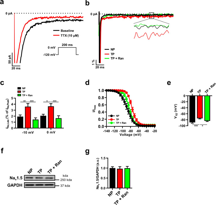
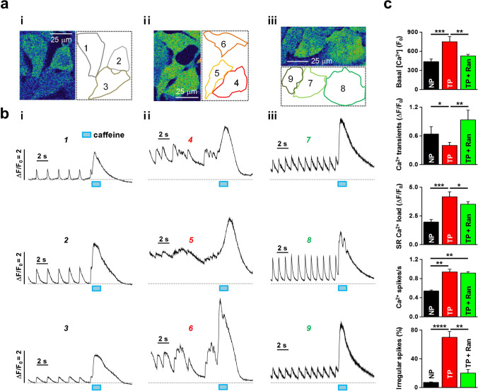
An aberrant late sodium current (I) caused by a mutation in the cardiac sodium channel (Na1.5) has emerged as a contributor to electrical remodeling that causes susceptibility to atrial fibrillation (AF). Although downregulation of phosphoinositide 3-kinase (PI3K)/Akt signaling is associated with AF, the molecular mechanisms underlying the negative regulation of I in AF remain unclear, and potential therapeutic approaches are needed. In this work, we constructed a tachypacing-induced cellular model of AF by exposing HL-1 myocytes to rapid electrical stimulation (1.5 V/cm, 4 ms, 10 Hz) for 6 h. Then, we gathered data using confocal Ca imaging, immunofluorescence, patch-clamp recordings, and immunoblots. The tachypacing cells displayed irregular Ca release, delayed afterdepolarization, prolonged action potential duration, and reduced PI3K/Akt signaling compared with controls. Those detrimental effects were related to increased I and were significantly mediated by treatment with the I blocker ranolazine. Furthermore, decreased PI3K/Akt signaling via PI3K inhibition increased I and subsequent aberrant myocyte excitability, which were abolished by I inhibition, suggesting that PI3K/Akt signaling is responsible for regulating pathogenic I. These results indicate that PI3K/Akt signaling is critical for regulating I and electrical remodeling, supporting the use of PI3K/Akt-mediated I as a therapeutic target for AF.
异常的晚钠电流 (I) 是由心脏钠通道 (Na1.5) 突变引起的,它已成为导致心房颤动 (AF) 易感性的电重构的一个因素。尽管磷酸肌醇 3-激酶 (PI3K)/Akt 信号转导的下调与 AF 相关,但 AF 中 I 的负调控的分子机制尚不清楚,需要潜在的治疗方法。在这项工作中,我们通过用 1.5 V/cm、4 ms、10 Hz 的快速电刺激暴露 HL-1 心肌细胞 6 小时来构建 AF 的电起搏诱导的细胞模型。然后,我们使用共聚焦 Ca 成像、免疫荧光、膜片钳记录和免疫印迹收集数据。与对照组相比,电起搏细胞显示不规则的 Ca 释放、延迟后除极、动作电位持续时间延长和 PI3K/Akt 信号转导减少。这些有害影响与 I 的增加有关,并且通过用 I 阻滞剂雷诺嗪处理可显著介导。此外,通过 PI3K 抑制降低 PI3K/Akt 信号转导可增加 I 和随后的异常心肌细胞兴奋性,而 I 抑制可消除这些作用,表明 PI3K/Akt 信号转导负责调节致病 I。这些结果表明,PI3K/Akt 信号转导对于调节 I 和电重构至关重要,支持将 PI3K/Akt 介导的 I 作为 AF 的治疗靶点。