• 文献检索
  • 文档翻译
  • 深度研究
  • 学术资讯
  • Suppr Zotero 插件Zotero 插件
  • 邀请有礼
  • 套餐&价格
  • 历史记录
应用&插件
Suppr Zotero 插件Zotero 插件浏览器插件Mac 客户端Windows 客户端微信小程序
定价
高级版会员购买积分包购买API积分包
服务
文献检索文档翻译深度研究API 文档MCP 服务
关于我们
关于 Suppr公司介绍联系我们用户协议隐私条款
关注我们

Suppr 超能文献

核心技术专利:CN118964589B侵权必究
粤ICP备2023148730 号-1Suppr @ 2026

文献检索

告别复杂PubMed语法,用中文像聊天一样搜索,搜遍4000万医学文献。AI智能推荐,让科研检索更轻松。

立即免费搜索

文件翻译

保留排版,准确专业,支持PDF/Word/PPT等文件格式,支持 12+语言互译。

免费翻译文档

深度研究

AI帮你快速写综述,25分钟生成高质量综述,智能提取关键信息,辅助科研写作。

立即免费体验

CT 血管造影在急性胃肠道出血中的作用:活跃和隐匿性发现的影像学描述。

The Role of CT-Angiography in the Acute Gastrointestinal Bleeding: A Pictorial Essay of Active and Obscure Findings.

机构信息

Department of General and Emergency Radiology, "Antonio Cardarelli" Hospital, Antonio Cardarelli St. 9, 80131 Naples, Italy.

Department of Gastroenterology and Digestive Endoscopy, "Antonio Cardarelli" Hospital, Antonio Cardarelli St. 9, 80131 Naples, Italy.

出版信息

Tomography. 2022 Sep 23;8(5):2369-2402. doi: 10.3390/tomography8050198.

DOI:10.3390/tomography8050198
PMID:36287797
原文链接:https://pmc.ncbi.nlm.nih.gov/articles/PMC9606936/
Abstract

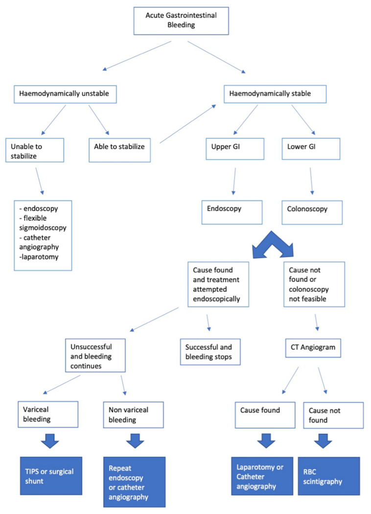
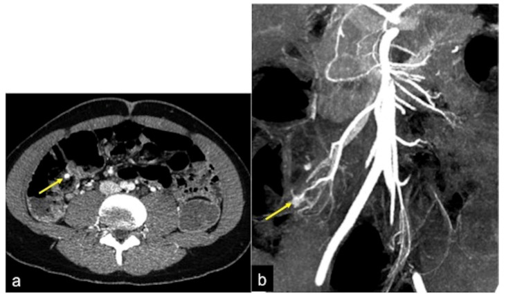
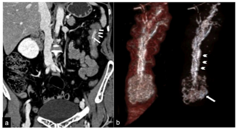
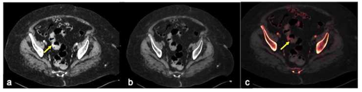
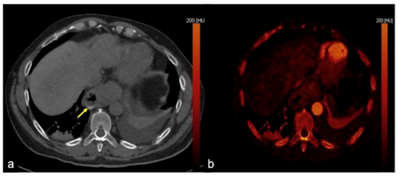
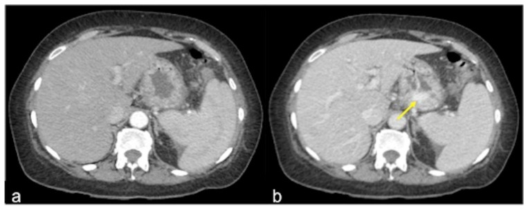
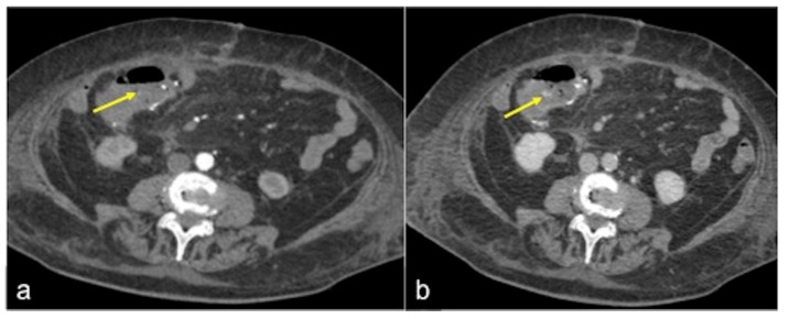
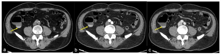
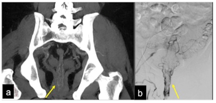
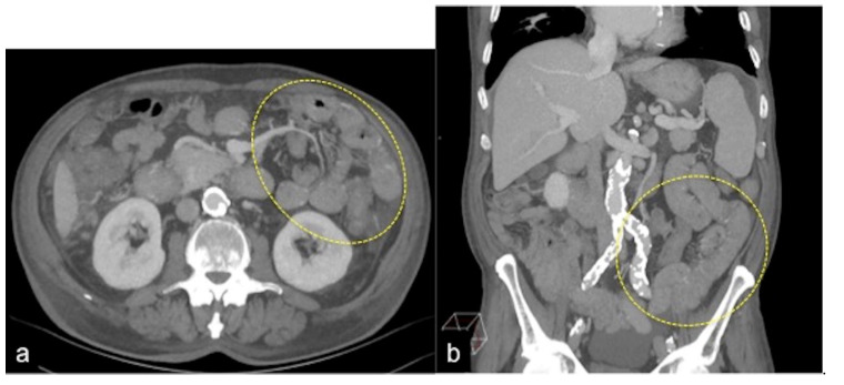
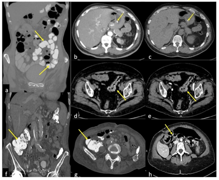
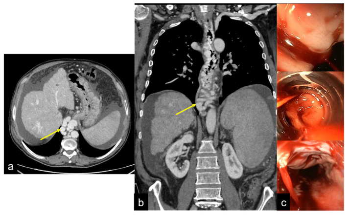
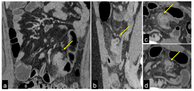
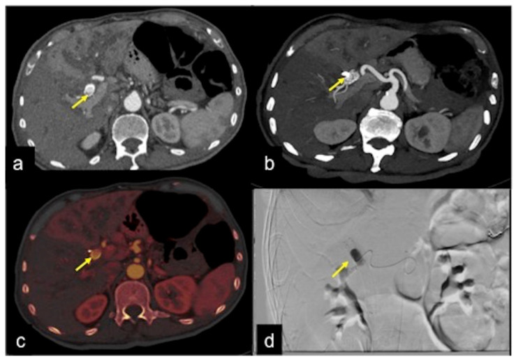
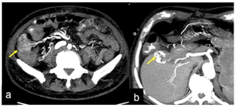
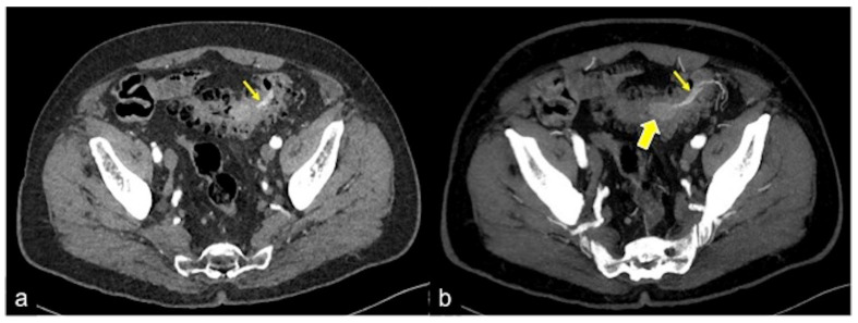
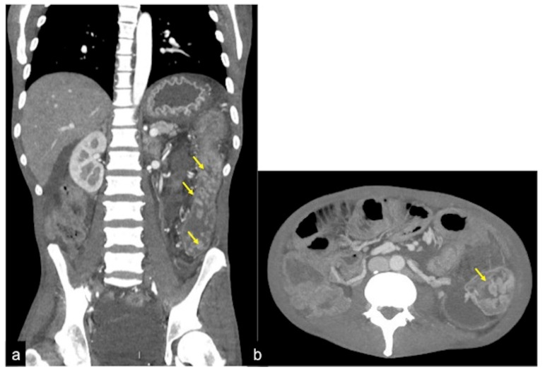
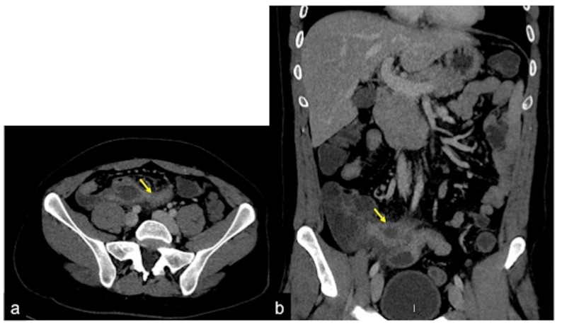
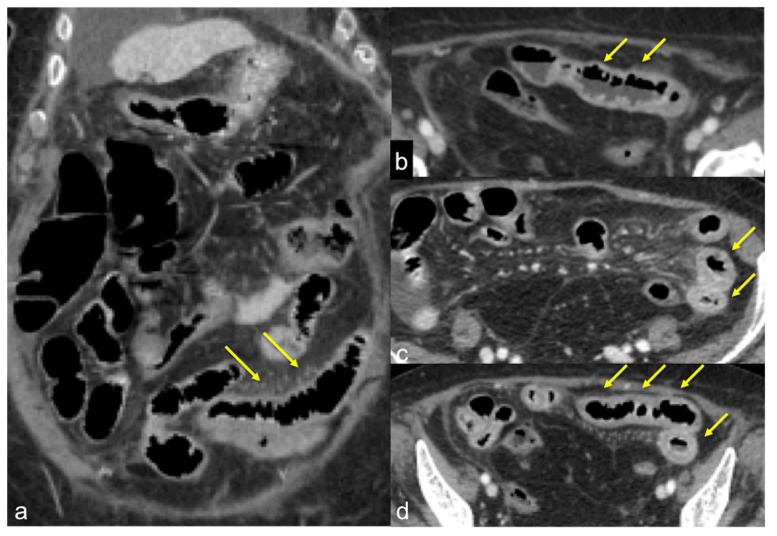
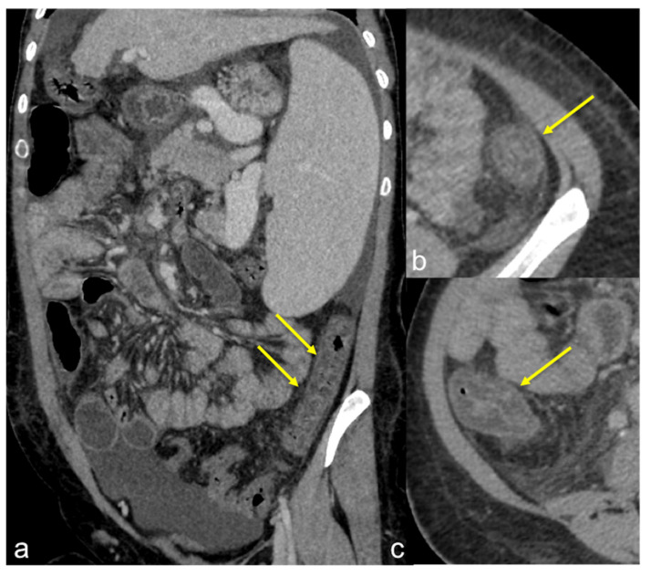
Gastrointestinal bleeding is a potentially life-threatening abdominal emergency that remains a common cause of hospitalisation. Although 80-85% of cases of gastrointestinal bleeding resolve spontaneously, it can result in massive haemorrhage and death. The presentation of gastrointestinal bleeding can range from asymptomatic or mildly ill patients requiring only conservative treatments to severely ill patients requiring immediate intervention. Identifying the source of the bleeding can be difficult due to the wide range of potential causes, the length of the gastrointestinal tract and the intermittent nature of the bleeding. The diagnostic and therapeutic approach is fully dependent on the nature of the bleeding and the patient's haemodynamic status. Radiologists should be aware of the appropriate uses of computed tomography angiography and other imaging modalities in patients with acute gastrointestinal bleeding, as well as the semiotics of bleeding and diagnostic pitfalls in order to appropriately diagnose and manage these patients. The learning objective of this review is to illustrate the computed tomography angiography technique, including the potential role of dual-energy computed tomography angiography, also highlighting the tips and tricks to identify the most common and uncommon features of acute gastrointestinal bleeding and its obscure form.

摘要

胃肠道出血是一种潜在的危及生命的腹部急症,仍然是导致住院的常见原因。尽管 80-85%的胃肠道出血病例会自发缓解,但它可能导致大量出血和死亡。胃肠道出血的表现范围从无症状或轻度不适的患者仅需保守治疗到需要立即干预的严重疾病患者。由于潜在原因广泛、胃肠道长度长以及出血间歇性,确定出血部位可能很困难。诊断和治疗方法完全取决于出血的性质和患者的血流动力学状态。放射科医生应该了解在急性胃肠道出血患者中使用计算机断层血管造影和其他成像方式的适当用途,以及出血的征象和诊断陷阱,以便对这些患者进行适当的诊断和治疗。本次综述的学习目标是说明计算机断层血管造影技术,包括双能计算机断层血管造影的潜在作用,还强调了识别急性胃肠道出血及其隐匿形式的最常见和不常见特征的技巧和窍门。

https://cdn.ncbi.nlm.nih.gov/pmc/blobs/605f/9606936/942ad7487d56/tomography-08-00198-g042.jpg
https://cdn.ncbi.nlm.nih.gov/pmc/blobs/605f/9606936/5f5740d1ea2e/tomography-08-00198-g001.jpg
https://cdn.ncbi.nlm.nih.gov/pmc/blobs/605f/9606936/c236dbae0c68/tomography-08-00198-g002.jpg
https://cdn.ncbi.nlm.nih.gov/pmc/blobs/605f/9606936/b687a33249de/tomography-08-00198-g004.jpg
https://cdn.ncbi.nlm.nih.gov/pmc/blobs/605f/9606936/59f621ad868d/tomography-08-00198-g005.jpg
https://cdn.ncbi.nlm.nih.gov/pmc/blobs/605f/9606936/00b6dc688417/tomography-08-00198-g006.jpg
https://cdn.ncbi.nlm.nih.gov/pmc/blobs/605f/9606936/00507ca15806/tomography-08-00198-g007.jpg
https://cdn.ncbi.nlm.nih.gov/pmc/blobs/605f/9606936/f71c317afd3a/tomography-08-00198-g008.jpg
https://cdn.ncbi.nlm.nih.gov/pmc/blobs/605f/9606936/38f86c6dbe94/tomography-08-00198-g009.jpg
https://cdn.ncbi.nlm.nih.gov/pmc/blobs/605f/9606936/6f621a05ba4b/tomography-08-00198-g010.jpg
https://cdn.ncbi.nlm.nih.gov/pmc/blobs/605f/9606936/13bc08047c1e/tomography-08-00198-g011.jpg
https://cdn.ncbi.nlm.nih.gov/pmc/blobs/605f/9606936/57212e939e03/tomography-08-00198-g012.jpg
https://cdn.ncbi.nlm.nih.gov/pmc/blobs/605f/9606936/ee0a6a977350/tomography-08-00198-g013.jpg
https://cdn.ncbi.nlm.nih.gov/pmc/blobs/605f/9606936/9cbf290d42be/tomography-08-00198-g014.jpg
https://cdn.ncbi.nlm.nih.gov/pmc/blobs/605f/9606936/174f303faf13/tomography-08-00198-g015.jpg
https://cdn.ncbi.nlm.nih.gov/pmc/blobs/605f/9606936/c0cd2b0f42dc/tomography-08-00198-g016.jpg
https://cdn.ncbi.nlm.nih.gov/pmc/blobs/605f/9606936/5dbb85b69cfb/tomography-08-00198-g017.jpg
https://cdn.ncbi.nlm.nih.gov/pmc/blobs/605f/9606936/1bd2421c2456/tomography-08-00198-g018.jpg
https://cdn.ncbi.nlm.nih.gov/pmc/blobs/605f/9606936/ed68bf4650db/tomography-08-00198-g019.jpg
https://cdn.ncbi.nlm.nih.gov/pmc/blobs/605f/9606936/8d74fcd7446c/tomography-08-00198-g021.jpg
https://cdn.ncbi.nlm.nih.gov/pmc/blobs/605f/9606936/253207f73151/tomography-08-00198-g030.jpg
https://cdn.ncbi.nlm.nih.gov/pmc/blobs/605f/9606936/83182188a096/tomography-08-00198-g032.jpg
https://cdn.ncbi.nlm.nih.gov/pmc/blobs/605f/9606936/0bb3428e741f/tomography-08-00198-g034.jpg
https://cdn.ncbi.nlm.nih.gov/pmc/blobs/605f/9606936/af6e76c55d1b/tomography-08-00198-g036.jpg
https://cdn.ncbi.nlm.nih.gov/pmc/blobs/605f/9606936/26720967f847/tomography-08-00198-g037.jpg
https://cdn.ncbi.nlm.nih.gov/pmc/blobs/605f/9606936/f0ec79934841/tomography-08-00198-g038.jpg
https://cdn.ncbi.nlm.nih.gov/pmc/blobs/605f/9606936/2a2614639815/tomography-08-00198-g039.jpg
https://cdn.ncbi.nlm.nih.gov/pmc/blobs/605f/9606936/2b0388ef1294/tomography-08-00198-g040.jpg
https://cdn.ncbi.nlm.nih.gov/pmc/blobs/605f/9606936/11ab1ef16904/tomography-08-00198-g041.jpg
https://cdn.ncbi.nlm.nih.gov/pmc/blobs/605f/9606936/942ad7487d56/tomography-08-00198-g042.jpg

相似文献

1
The Role of CT-Angiography in the Acute Gastrointestinal Bleeding: A Pictorial Essay of Active and Obscure Findings.CT 血管造影在急性胃肠道出血中的作用:活跃和隐匿性发现的影像学描述。
Tomography. 2022 Sep 23;8(5):2369-2402. doi: 10.3390/tomography8050198.
2
Acute gastrointestinal bleeding: emerging role of multidetector CT angiography and review of current imaging techniques.急性胃肠道出血:多排CT血管造影的新作用及当前成像技术综述
Radiographics. 2007 Jul-Aug;27(4):1055-70. doi: 10.1148/rg.274065095.
3
Gastrointestinal Bleeding at CT Angiography and CT Enterography: Imaging Atlas and Glossary of Terms.CT 血管造影和 CT 肠造影中的胃肠道出血:成像图谱和术语词汇表。
Radiographics. 2021 Oct;41(6):1632-1656. doi: 10.1148/rg.2021210043.
4
The role of computerized tomography in the evaluation of gastrointestinal bleeding following negative or failed endoscopy: a review of current status.计算机断层扫描在评估内镜检查阴性或失败后的胃肠道出血中的作用:现状综述
J Postgrad Med. 2008 Apr-Jun;54(2):126-34. doi: 10.4103/0022-3859.40779.
5
Diagnostic Performance of CT Angiography in Patients Visiting Emergency Department with Overt Gastrointestinal Bleeding.CT血管造影术对急诊就诊的显性胃肠道出血患者的诊断效能
Korean J Radiol. 2015 May-Jun;16(3):541-9. doi: 10.3348/kjr.2015.16.3.541. Epub 2015 May 13.
6
Acute lower intestinal bleeding: feasibility and diagnostic performance of CT angiography.急性下消化道出血:CT 血管造影的可行性和诊断性能。
Radiology. 2012 Jan;262(1):109-16. doi: 10.1148/radiol.11110326. Epub 2011 Nov 14.
7
CT for Evaluation of Acute Gastrointestinal Bleeding.CT 用于急性胃肠道出血的评估。
Radiographics. 2018 Jul-Aug;38(4):1089-1107. doi: 10.1148/rg.2018170138. Epub 2018 Jun 8.
8
Multidetector CT angiography in acute gastrointestinal bleeding: why, when, and how.多排 CT 血管造影在急性胃肠道出血中的应用:为何、何时及如何应用。
Radiographics. 2011 May-Jun;31(3):E35-46. doi: 10.1148/rg.313105206.
9
Acute gastrointestinal bleeding: CT angiography with multi-planar reformatting.急性胃肠道出血:采用多平面重组的CT血管造影术
Abdom Imaging. 2011 Apr;36(2):115-25. doi: 10.1007/s00261-010-9615-7.
10
[Massive small intestine bleeding: CT-angiography and surgical treatment - a case report].[大量小肠出血:CT血管造影及手术治疗——病例报告]
Rozhl Chir. 2015 Apr;94(4):174-6.

引用本文的文献

1
Endovascular Localization of Aortic Injury in a Porcine Model.猪模型中主动脉损伤的血管内定位
IEEE Open J Eng Med Biol. 2025 Apr 2;6:425-431. doi: 10.1109/OJEMB.2025.3556987. eCollection 2025.
2
Successful Management of Acute Obscure Gastrointestinal Bleeding (OGIB) Using Long Tube-Directed Hemostasis: A Case Report.长管引导下止血成功治疗急性不明原因消化道出血(OGIB):病例报告
Cureus. 2025 May 16;17(5):e84251. doi: 10.7759/cureus.84251. eCollection 2025 May.
3
Transarterial embolization for acute lower gastrointestinal bleeding: a retrospective bicentric study.

本文引用的文献

1
Gastrointestinal Bleeding at CT Angiography and CT Enterography: Imaging Atlas and Glossary of Terms.CT 血管造影和 CT 肠造影中的胃肠道出血:成像图谱和术语词汇表。
Radiographics. 2021 Oct;41(6):1632-1656. doi: 10.1148/rg.2021210043.
2
Computed tomography imaging of septic shock. Beyond the cause: the "CT hypoperfusion complex". A pictorial essay.脓毒性休克的计算机断层扫描成像。病因之外:“CT灌注不足复合体”。一篇图文并茂的文章。
Insights Imaging. 2021 Jun 5;12(1):70. doi: 10.1186/s13244-021-01006-5.
3
Large Bowel Ischemia/Infarction: How to Recognize It and Make Differential Diagnosis? A Review.
经动脉栓塞治疗急性下消化道出血:一项回顾性双中心研究
Radiol Med. 2025 Apr 18. doi: 10.1007/s11547-025-02012-z.
4
A Rare Encounter: Gastric Ulcer Penetration into the Splenic Hilum Presenting with Upper Gastrointestinal Bleeding and a Massive Splenic Haematoma-Case Report and Literature Review.罕见病例:胃溃疡穿透至脾门并伴有上消化道出血及巨大脾血肿——病例报告与文献综述
Diagnostics (Basel). 2025 Mar 4;15(5):617. doi: 10.3390/diagnostics15050617.
5
Empiric embolization by vasospasm therapy for acute lower gastrointestinal bleeding: a preliminary report.经验性血管痉挛治疗栓塞术治疗急性下消化道出血:初步报告。
Sci Rep. 2024 Oct 28;14(1):25728. doi: 10.1038/s41598-024-76408-8.
6
Extravasated contrast volumetric assessment on computed tomography angiography in gastrointestinal bleeding: A useful predictor of positive angiographic findings.胃肠道出血计算机断层血管造影中造影剂外渗的容积评估:血管造影阳性结果的有用预测指标。
World J Radiol. 2024 May 28;16(5):115-127. doi: 10.4329/wjr.v16.i5.115.
7
Artificial Intelligence for Identification of Images with Active Bleeding in Mesenteric and Celiac Arteries Angiography.人工智能在肠系膜和腹腔动脉血管造影中识别活动性出血的图像。
Cardiovasc Intervent Radiol. 2024 Jun;47(6):785-792. doi: 10.1007/s00270-024-03689-x. Epub 2024 Mar 26.
8
Value of multidetector computed tomography angiography in severe non-variceal upper gastrointestinal bleeding: a retrospective study in a referral bleeding unit.多排螺旋 CT 血管造影在重症非静脉曲张性上消化道出血中的价值:在一个转诊出血中心的回顾性研究。
Abdom Radiol (NY). 2024 May;49(5):1385-1396. doi: 10.1007/s00261-024-04208-9. Epub 2024 Mar 4.
9
Rare causes of acute non-variceal upper gastrointestinal bleeding: A comprehensive review.急性非静脉曲张性上消化道出血的罕见病因:全面综述。
World J Gastroenterol. 2023 Jul 21;29(27):4222-4235. doi: 10.3748/wjg.v29.i27.4222.
10
Practical Aspects of Upper Gastrointestinal Bleeding in Children.儿童上消化道出血的实际问题
J Clin Med. 2023 Apr 17;12(8):2921. doi: 10.3390/jcm12082921.
大肠缺血/梗死:如何识别及进行鉴别诊断?综述
Diagnostics (Basel). 2021 May 30;11(6):998. doi: 10.3390/diagnostics11060998.
4
Endoscopic diagnosis and management of nonvariceal upper gastrointestinal hemorrhage (NVUGIH): European Society of Gastrointestinal Endoscopy (ESGE) Guideline - Update 2021.内镜诊断和非静脉曲张性上消化道出血(NVUGIH)的处理:欧洲胃肠道内镜学会(ESGE)指南 - 2021 年更新。
Endoscopy. 2021 Mar;53(3):300-332. doi: 10.1055/a-1369-5274. Epub 2021 Feb 10.
5
Primary aortogastric fistula: an extraordinary rare endoscopic finding in the setting of upper gastrointestinal bleeding.原发性主动脉胃瘘:上消化道出血情况下一种极其罕见的内镜检查发现。
Endoscopy. 2021 Feb;53(2):E60-E61. doi: 10.1055/a-1180-7701. Epub 2020 Jun 19.
6
Dual-energy CT evaluation of gastrointestinal bleeding.双能 CT 评价胃肠道出血。
Abdom Radiol (NY). 2020 Jan;45(1):1-14. doi: 10.1007/s00261-019-02226-6.
7
The role of imaging in gastrointestinal bleed.影像学在胃肠道出血中的作用。
Cardiovasc Diagn Ther. 2019 Aug;9(Suppl 1):S88-S96. doi: 10.21037/cdt.2018.12.07.
8
[Drug-induced gastrointestinal bleeding].[药物性胃肠道出血]
Internist (Berl). 2019 Jun;60(6):597-607. doi: 10.1007/s00108-019-0610-y.
9
The pediatric gastrointestinal tract: ultrasound findings in acute diseases.小儿胃肠道:急性疾病的超声表现
J Ultrasound. 2019 Dec;22(4):409-422. doi: 10.1007/s40477-018-00355-0. Epub 2019 Feb 13.
10
MDCT in acute ischaemic left colitis: a pictorial essay.MDCT 检查在急性缺血性左结肠炎中的应用:影像学研究。
Radiol Med. 2019 Feb;124(2):103-108. doi: 10.1007/s11547-018-0947-7. Epub 2018 Oct 27.