Di Serafino Marco, Viscardi Daniela, Iacobellis Francesca, Giugliano Luigi, Barbuto Luigi, Oliva Gaspare, Ronza Roberto, Borzelli Antonio, Raucci Antonio, Pezzullo Filomena, De Cristofaro Maria Giovanna, Romano Luigia
Department of General and Emergency Radiology, "Antonio Cardarelli" Hospital, Antonio Cardarelli st 9, 80131, Naples, Italy.
Department of Anesthesia and Resuscitation, "Antonio Cardarelli" Hospital, Naples, Italy.
Insights Imaging. 2021 Jun 5;12(1):70. doi: 10.1186/s13244-021-01006-5.
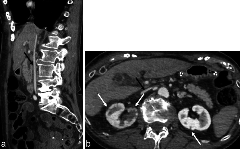
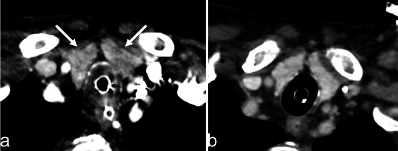
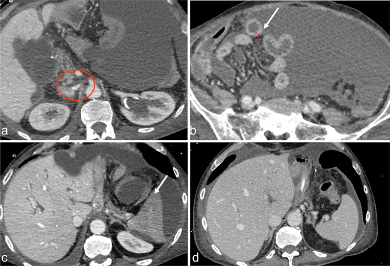
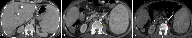
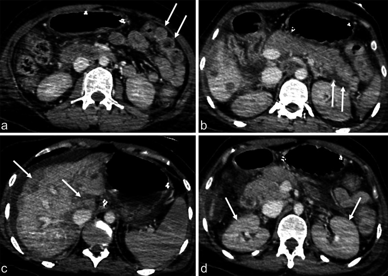
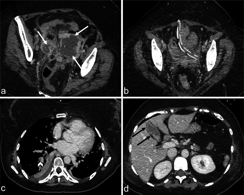
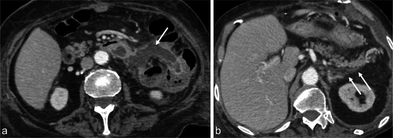
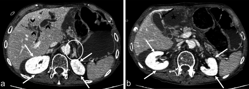
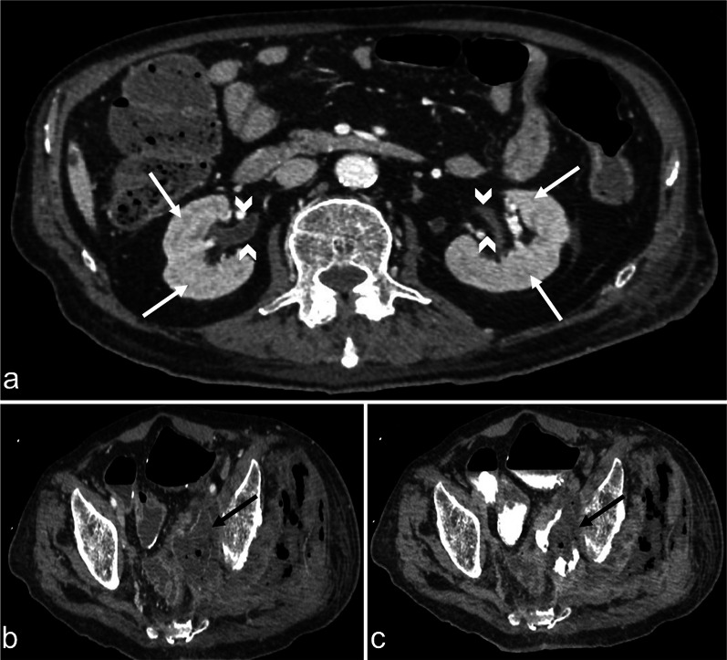
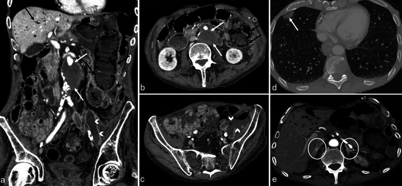
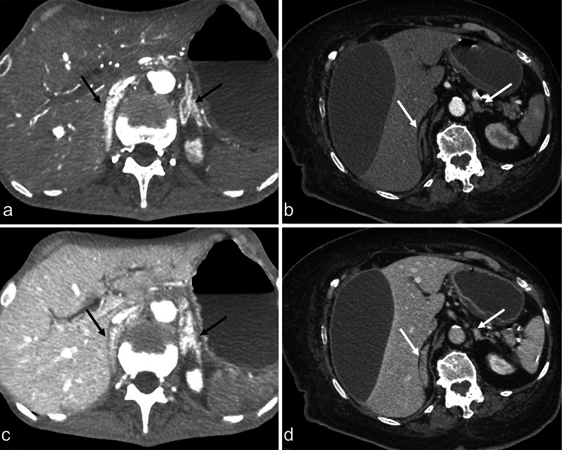
Septic shock is a medical emergency that represents one of the most important underlying causes for presentation to the Emergency Department. Sepsis is defined as organ dysfunction, life-threatening event caused by a deregulated inflammatory host response to infection, with a mortality risk ranging from 10 to 40%. Early sepsis identification is the cornerstone of management and diagnostic imaging can play a pivotal role in this clinical context. The choice of imaging modality depends on several factors, associated with the clinical condition and the presence or absence of localising signs and symptoms. The diagnostic accuracy of contrast-enhanced total-body CT has been well established during septic shock, allowing for a rapid, panoramic, and detailed study of multiple body areas, simultaneously. The aim of this article is to illustrate the controversial CT hypoperfusion complex in patients with septic shock, characterised by the following imaging features: decreased enhancement of the viscera; increased mucosal enhancement; luminal dilation of the small bowel; mural thickening and fluid-filled loops of the small bowel; the halo sign and flattening of the inferior vena cava; reduced aortic diameter; peripancreatic oedema; abnormal parenchymal perfusion; and other controversial findings that are variably associated with each other and reversible during the early stages. Increasing physicians' awareness of the significance of these findings could prompt alternative approaches to the early assessment and management of septic shock. In this perspective, CT imaging represents a useful tool for a complete, rapid and detailed diagnosis of clinically suspected septic shock, which can be used to improve patient outcomes.
脓毒性休克是一种医疗急症,是患者前往急诊科就诊的最重要潜在病因之一。脓毒症被定义为由宿主对感染的炎症反应失调引起的器官功能障碍和危及生命的事件,死亡率在10%至40%之间。早期识别脓毒症是治疗的基石,而诊断性影像学检查在这一临床背景中可发挥关键作用。成像方式的选择取决于几个因素,与临床状况以及是否存在定位体征和症状有关。在脓毒性休克期间,对比增强全身CT的诊断准确性已得到充分证实,它能够同时对多个身体部位进行快速、全景且详细的检查。本文旨在阐述脓毒性休克患者中存在争议的CT灌注不足复合体,其具有以下影像学特征:内脏强化减弱;黏膜强化增加;小肠肠腔扩张;小肠壁增厚和肠袢积液;晕征以及下腔静脉变平;主动脉直径减小;胰腺周围水肿;实质灌注异常;以及其他一些存在争议的表现,这些表现相互之间存在不同程度的关联,且在早期阶段是可逆的。提高医生对这些发现重要性的认识可能会促使在脓毒性休克的早期评估和管理中采用不同的方法。从这个角度来看,CT成像对于临床疑似脓毒性休克的全面、快速和详细诊断是一种有用的工具,可用于改善患者的预后。