Parisi Francesca, Abramo Francesca, Maimone Marco, Poli Alessandro, Millanta Francesca
Department of Veterinary Sciences, University of Pisa, Viale delle Piagge n. 2, 56124 Pisa, Italy.
Clinica Veterinaria Foce, via Eugenio Baroni, 26R, 16129 Genova, Italy.
Vet Sci. 2022 Oct 5;9(10):548. doi: 10.3390/vetsci9100548.
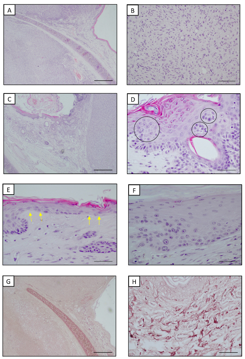
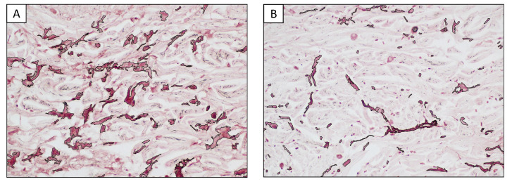
As with human species, recent studies also suggest a photoinduced etiopathology for non-epithelial cutaneous tumors in feline species. We report a recent case of a ten-year-old male cat with a white-hair coat and mesenchymal neoplasms of both auricles. Cytology, complete blood count (CBC), serum biochemistry and imaging examinations were performed. After surgery, the samples underwent routinary histopathology and were additionally stained with orcein. A routine analysis yielded values within a normal range and the imaging examination showed no abnormalities, suggesting that the bilateral presentation of neoplasms was primary rather than metastatic. The cytology was inconclusive, but, through histopathology, two well-differentiated fibrosarcomas were diagnosed and histopathological changes related to chronic UV exposure (such as epidermal hyperplasia, stratification disorders, keratinocyte dysplasia and an accumulation of elastotic material) were documented in the skin adjacent to the lesions. An orcein stain succeeded in highlighting elastosis. The elastic fibers lost their regular structure and orientation and appeared to be fragmented, wavy to branched and knotted. A morphometric analysis showed that the amount of elastotic material in the dermis close to the tumors was more than double compared with the more distant areas. Elastosis is considered to be a hallmark of photodamage; thus, an involvement of UV rays in the carcinogenic process of the tumors may be suspected.
与人类一样,最近的研究也表明猫科动物的非上皮性皮肤肿瘤存在光诱导病因学。我们报告了一例近期病例,一只十岁雄性白猫双耳患有间质性肿瘤。进行了细胞学检查、全血细胞计数(CBC)、血清生化检查和影像学检查。手术后,样本进行了常规组织病理学检查,并额外用orcein染色。常规分析结果在正常范围内,影像学检查未发现异常,表明肿瘤的双侧表现是原发性的而非转移性的。细胞学检查结果不明确,但通过组织病理学检查,诊断出两个高分化纤维肉瘤,并且在病变附近的皮肤中记录到了与慢性紫外线暴露相关的组织病理学变化(如表皮增生、分层紊乱、角质形成细胞发育异常和弹性物质积聚)。orcein染色成功地突出了弹性组织变性。弹性纤维失去了其规则的结构和方向,看起来破碎、呈波浪状至分支状且打结。形态计量分析表明,靠近肿瘤的真皮中弹性物质的量是较远区域的两倍多。弹性组织变性被认为是光损伤的标志;因此,可能怀疑紫外线参与了肿瘤的致癌过程。