• 文献检索
  • 文档翻译
  • 深度研究
  • 学术资讯
  • Suppr Zotero 插件Zotero 插件
  • 邀请有礼
  • 套餐&价格
  • 历史记录
应用&插件
Suppr Zotero 插件Zotero 插件浏览器插件Mac 客户端Windows 客户端微信小程序
定价
高级版会员购买积分包购买API积分包
服务
文献检索文档翻译深度研究API 文档MCP 服务
关于我们
关于 Suppr公司介绍联系我们用户协议隐私条款
关注我们

Suppr 超能文献

核心技术专利:CN118964589B侵权必究
粤ICP备2023148730 号-1Suppr @ 2026

文献检索

告别复杂PubMed语法,用中文像聊天一样搜索,搜遍4000万医学文献。AI智能推荐,让科研检索更轻松。

立即免费搜索

文件翻译

保留排版,准确专业,支持PDF/Word/PPT等文件格式,支持 12+语言互译。

免费翻译文档

深度研究

AI帮你快速写综述,25分钟生成高质量综述,智能提取关键信息,辅助科研写作。

立即免费体验

PPARα 的激活可改善 Dsg2 缺陷型心律失常性心肌病的心肌纤维化。

Activation of PPARα Ameliorates Cardiac Fibrosis in Dsg2-Deficient Arrhythmogenic Cardiomyopathy.

机构信息

Department of Physiology, School of Medicine, Jinan University, 601 Huangpu Avenue West, Tianhe District, Guangzhou 510632, China.

Center for Clinical Epidemiology and Methodology (CCEM), Guangdong Second Provincial General Hospital, Guangzhou 510317, China.

出版信息

Cells. 2022 Oct 11;11(20):3184. doi: 10.3390/cells11203184.

DOI:10.3390/cells11203184
PMID:36291052
原文链接:https://pmc.ncbi.nlm.nih.gov/articles/PMC9601208/
Abstract

BACKGROUND

Arrhythmogenic cardiomyopathy (ACM) is a genetic heart muscle disease characterized by progressive fibro-fatty replacement of cardiac myocytes. Up to now, the existing therapeutic modalities for ACM are mostly palliative. About 50% of ACM is caused by mutations in genes encoding desmosomal proteins including Desmoglein-2 (Dsg2). In the current study, the cardiac fibrosis of ACM and its underlying mechanism were investigated by using a cardiac-specific knockout of Dsg2 mouse model.

METHODS

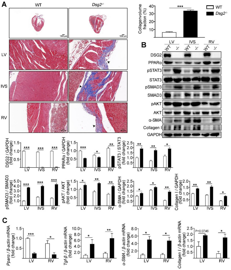
Cardiac-specific knockout (CS-Dsg2) mice and wild-type (WT) mice were respectively used as the animal model of ACM and controls. The myocardial collagen volume fraction was determined by histological analysis. The expression levels of fibrotic markers such as α-SMA and Collagen I as well as signal transducers such as STAT3, SMAD3, and PPARα were measured by Western blot and quantitative real-time PCR.

RESULTS

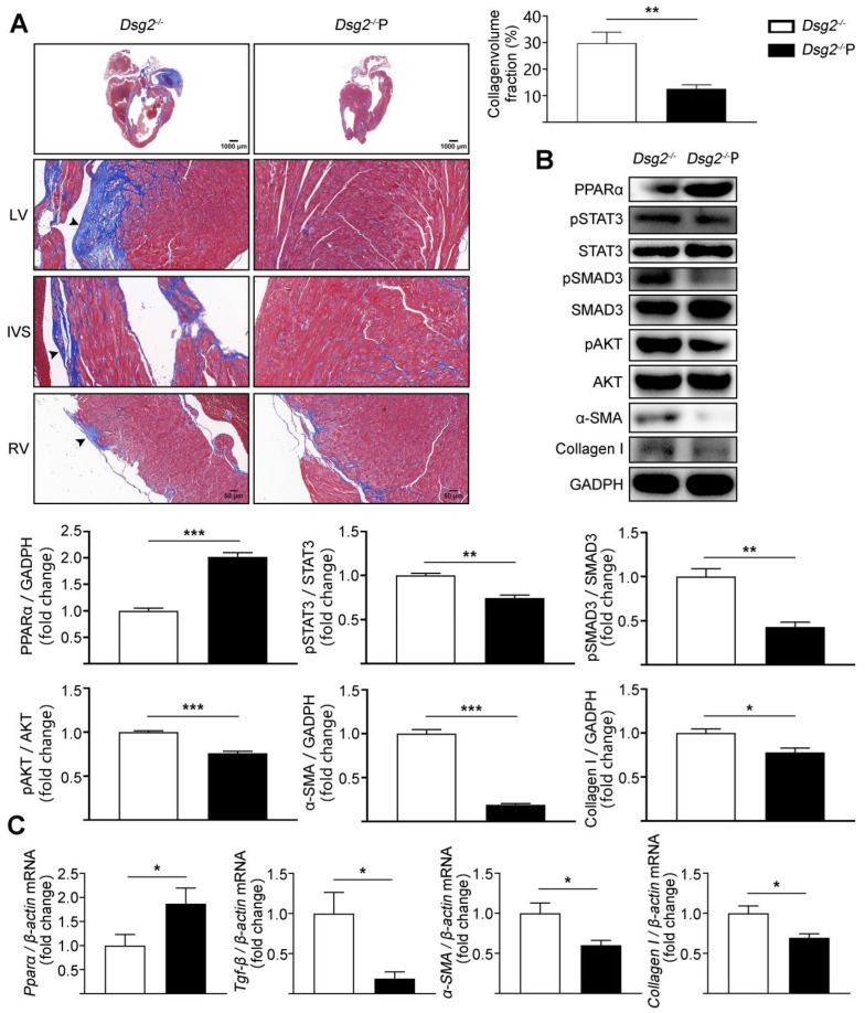
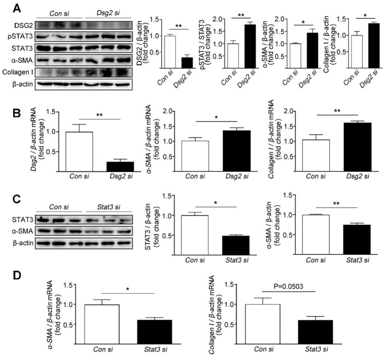
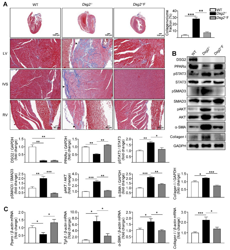
Increased cardiac fibrosis was observed in CS-Dsg2 mice according to Masson staining. PPARα deficiency and hyperactivation of STAT3 and SMAD3 were observed in the myocardium of CS-Dsg2 mice. The biomarkers of fibrosis such as α-SMA and Collagen I were upregulated after gene silencing of Dsg2 in HL-1 cells. Furthermore, STAT3 gene silencing by Stat3 siRNA inhibited the expression of fibrotic markers. The activation of PPARα by fenofibrate or AAV9-Pparα improved the cardiac fibrosis and decreased the phosphorylation of STAT3, SMAD3, and AKT in CS-Dsg2 mice.

CONCLUSIONS

Activation of PPARα alleviates the cardiac fibrosis in ACM.

摘要

背景

致心律失常性右室心肌病(ACM)是一种遗传性心肌疾病,其特征是心肌细胞进行性纤维脂肪替代。到目前为止,ACM 的现有治疗方法大多是姑息性的。大约 50%的 ACM 是由桥粒蛋白编码基因(包括桥粒芯糖蛋白 2 [Dsg2])的突变引起的。在本研究中,通过使用心脏特异性 Dsg2 敲除小鼠模型研究了 ACM 的心脏纤维化及其潜在机制。

方法

心脏特异性敲除(CS-Dsg2)小鼠和野生型(WT)小鼠分别作为 ACM 的动物模型和对照。通过组织学分析确定心肌胶原容积分数。通过 Western blot 和定量实时 PCR 测量纤维化标志物(如α-SMA 和 Collagen I)和信号转导物(如 STAT3、SMAD3 和 PPARα)的表达水平。

结果

Masson 染色显示 CS-Dsg2 小鼠的心脏纤维化增加。CS-Dsg2 小鼠的心肌中观察到 PPARα 缺失和 STAT3 和 SMAD3 的过度激活。HL-1 细胞中 Dsg2 基因沉默后,纤维化标志物如α-SMA 和 Collagen I 的表达上调。此外,Stat3 siRNA 抑制 STAT3 基因沉默可抑制纤维化标志物的表达。非诺贝特或 AAV9-Pparα 激活 PPARα 可改善 CS-Dsg2 小鼠的心脏纤维化,并降低 CS-Dsg2 小鼠心脏中 STAT3、SMAD3 和 AKT 的磷酸化。

结论

激活 PPARα 可减轻 ACM 中的心脏纤维化。

https://cdn.ncbi.nlm.nih.gov/pmc/blobs/ca66/9601208/ca5ffcb97a53/cells-11-03184-g004.jpg
https://cdn.ncbi.nlm.nih.gov/pmc/blobs/ca66/9601208/36f0269f19e1/cells-11-03184-g001.jpg
https://cdn.ncbi.nlm.nih.gov/pmc/blobs/ca66/9601208/f57416e7c8ff/cells-11-03184-g002.jpg
https://cdn.ncbi.nlm.nih.gov/pmc/blobs/ca66/9601208/413b447ae061/cells-11-03184-g003.jpg
https://cdn.ncbi.nlm.nih.gov/pmc/blobs/ca66/9601208/ca5ffcb97a53/cells-11-03184-g004.jpg

相似文献

1
Activation of PPARα Ameliorates Cardiac Fibrosis in Dsg2-Deficient Arrhythmogenic Cardiomyopathy.PPARα 的激活可改善 Dsg2 缺陷型心律失常性心肌病的心肌纤维化。
Cells. 2022 Oct 11;11(20):3184. doi: 10.3390/cells11203184.
2
Hyperactivation of ATF4/TGF-β1 signaling contributes to the progressive cardiac fibrosis in Arrhythmogenic cardiomyopathy caused by DSG2 Variant.DSG2 变异导致心律失常性心肌病中 ATF4/TGF-β1 信号的过度激活导致进行性心肌纤维化。
BMC Med. 2024 Sep 4;22(1):361. doi: 10.1186/s12916-024-03593-8.
3
Reactivation of PPAR alleviates myocardial lipid accumulation and cardiac dysfunction by improving fatty acid -oxidation in -deficient arrhythmogenic cardiomyopathy.过氧化物酶体增殖物激活受体(PPAR)的激活通过改善致心律失常性心肌病中脂肪酸氧化的缺陷,减轻心肌脂质蓄积和心脏功能障碍。
Acta Pharm Sin B. 2023 Jan;13(1):192-203. doi: 10.1016/j.apsb.2022.05.018. Epub 2022 May 21.
4
Defective Desmosomal Adhesion Causes Arrhythmogenic Cardiomyopathy by Involving an Integrin-αVβ6/TGF-β Signaling Cascade.桥粒连接缺陷通过整合素-αVβ6/TGF-β信号级联导致心律失常性心肌病。
Circulation. 2022 Nov 22;146(21):1610-1626. doi: 10.1161/CIRCULATIONAHA.121.057329. Epub 2022 Oct 21.
5
Intercalated disc abnormalities, reduced Na(+) current density, and conduction slowing in desmoglein-2 mutant mice prior to cardiomyopathic changes.连接蛋白 2 突变小鼠在出现心肌病性改变之前出现连接盘异常、钠电流密度降低和传导减慢。
Cardiovasc Res. 2012 Sep 1;95(4):409-18. doi: 10.1093/cvr/cvs219. Epub 2012 Jul 3.
6
Desmoglein 2-Dependent Arrhythmogenic Cardiomyopathy Is Caused by a Loss of Adhesive Function.桥粒芯糖蛋白2相关致心律失常性心肌病由黏附功能丧失所致。
Circ Cardiovasc Genet. 2015 Aug;8(4):553-63. doi: 10.1161/CIRCGENETICS.114.000974. Epub 2015 Jun 17.
7
In vitro analysis of arrhythmogenic cardiomyopathy associated desmoglein-2 (DSG2) mutations reveals diverse glycosylation patterns.体外分析心律失常性心肌病相关桥粒芯糖蛋白-2(DSG2)突变揭示了不同的糖基化模式。
J Mol Cell Cardiol. 2019 Apr;129:303-313. doi: 10.1016/j.yjmcc.2019.03.014. Epub 2019 Mar 15.
8
High-Fat Diet Augments Myocardial Inflammation and Cardiac Dysfunction in Arrhythmogenic Cardiomyopathy.高脂饮食可加剧致心律失常性心肌病的心肌炎症和心功能障碍。
Nutrients. 2024 Jun 29;16(13):2087. doi: 10.3390/nu16132087.
9
Peroxisome proliferator-activated receptor alpha-independent actions of fenofibrate exacerbates left ventricular dilation and fibrosis in chronic pressure overload.非诺贝特不依赖过氧化物酶体增殖物激活受体α的作用会加重慢性压力超负荷下的左心室扩张和纤维化。
Hypertension. 2007 May;49(5):1084-94. doi: 10.1161/HYPERTENSIONAHA.107.086926. Epub 2007 Mar 12.
10
Ser194Leu DSG2 mutation, associated with arrhythmogenic left ventricular cardiomyopathy and ventricular tachycardia.DSG2 基因 Ser194Leu 突变,与致心律失常性左室心肌病和室性心动过速相关。
Pacing Clin Electrophysiol. 2024 Apr;47(4):503-510. doi: 10.1111/pace.14950. Epub 2024 Feb 20.

引用本文的文献

1
Fenofibrate prevents myocardial inflammation and fibrosis via PPARα/IκBζ signaling pathway in rat autoimmune myocarditis.非诺贝特通过PPARα/IκBζ信号通路预防大鼠自身免疫性心肌炎中的心肌炎症和纤维化。
Exp Ther Med. 2025 May 6;30(1):128. doi: 10.3892/etm.2025.12879. eCollection 2025 Jul.
2
SFRP4 Knockdown Attenuates Dsg2-Deficient Arrhythmogenic Cardiomyopathy by Down-Regulating TGF-β and Smad3.SFRP4基因敲低通过下调TGF-β和Smad3减轻Dsg2缺陷型致心律失常性心肌病
Biochem Genet. 2025 Feb 28. doi: 10.1007/s10528-025-11052-z.
3
In Vivo Approaches to Understand Arrhythmogenic Cardiomyopathy: Perspectives on Animal Models.

本文引用的文献

1
Reactivation of PPAR alleviates myocardial lipid accumulation and cardiac dysfunction by improving fatty acid -oxidation in -deficient arrhythmogenic cardiomyopathy.过氧化物酶体增殖物激活受体(PPAR)的激活通过改善致心律失常性心肌病中脂肪酸氧化的缺陷,减轻心肌脂质蓄积和心脏功能障碍。
Acta Pharm Sin B. 2023 Jan;13(1):192-203. doi: 10.1016/j.apsb.2022.05.018. Epub 2022 May 21.
2
DYRK1B-STAT3 Drives Cardiac Hypertrophy and Heart Failure by Impairing Mitochondrial Bioenergetics.DYRK1B-STAT3 通过损害线粒体生物能学驱动心肌肥厚和心力衰竭。
Circulation. 2022 Mar 15;145(11):829-846. doi: 10.1161/CIRCULATIONAHA.121.055727. Epub 2022 Mar 2.
3
在体方法理解致心律失常性心肌病:动物模型的观点。
Cells. 2024 Jul 27;13(15):1264. doi: 10.3390/cells13151264.
4
Animal Models and Molecular Pathogenesis of Arrhythmogenic Cardiomyopathy Associated with Pathogenic Variants in Intercalated Disc Genes.与闰盘基因致病性变异相关的心律失常性心肌病的动物模型和分子发病机制。
Int J Mol Sci. 2024 Jun 5;25(11):6208. doi: 10.3390/ijms25116208.
5
Management Strategies in Arrhythmogenic Cardiomyopathy across the Spectrum of Ventricular Involvement.致心律失常性心肌病全谱心室受累的管理策略
Biomedicines. 2023 Dec 9;11(12):3259. doi: 10.3390/biomedicines11123259.
6
Understanding Arrhythmogenic Cardiomyopathy: Advances through the Use of Human Pluripotent Stem Cell Models.了解致心律失常性心肌病:通过使用人类多能干细胞模型取得的进展。
Genes (Basel). 2023 Sep 25;14(10):1864. doi: 10.3390/genes14101864.
7
Bioactive Compounds and Cardiac Fibrosis: Current Insight and Future Prospect.生物活性化合物与心脏纤维化:当前见解与未来展望
J Cardiovasc Dev Dis. 2023 Jul 21;10(7):313. doi: 10.3390/jcdd10070313.
8
Recent Insights into the Role of PPARs in Disease.PPARs 在疾病中的作用的最新见解。
Cells. 2023 Jun 7;12(12):1572. doi: 10.3390/cells12121572.
9
Gene editing innovations and their applications in cardiomyopathy research.基因编辑创新及其在心肌病研究中的应用。
Dis Model Mech. 2023 May 1;16(5). doi: 10.1242/dmm.050088. Epub 2023 May 24.
10
Targeting lipid metabolism as a new therapeutic strategy for inherited cardiomyopathies.将脂质代谢作为遗传性心肌病的一种新治疗策略。
Front Cardiovasc Med. 2023 Jan 19;10:1114459. doi: 10.3389/fcvm.2023.1114459. eCollection 2023.
Histopathological Features and Protein Markers of Arrhythmogenic Cardiomyopathy.
致心律失常性心肌病的组织病理学特征和蛋白质标志物
Front Cardiovasc Med. 2021 Dec 7;8:746321. doi: 10.3389/fcvm.2021.746321. eCollection 2021.
4
Nanoparticle conjugation of ginsenoside Rb3 inhibits myocardial fibrosis by regulating PPARα pathway.纳米粒子结合人参皂苷 Rb3 通过调节 PPARα 通路抑制心肌纤维化。
Biomed Pharmacother. 2021 Jul;139:111630. doi: 10.1016/j.biopha.2021.111630. Epub 2021 May 1.
5
Regulatory T cells promote glioma cell stemness through TGF-β-NF-κB-IL6-STAT3 signaling.调节性 T 细胞通过 TGF-β-NF-κB-IL6-STAT3 信号通路促进神经胶质瘤细胞干性。
Cancer Immunol Immunother. 2021 Sep;70(9):2601-2616. doi: 10.1007/s00262-021-02872-0. Epub 2021 Feb 12.
6
Emerging therapeutic targets for cardiac arrhythmias: role of STAT3 in regulating cardiac fibroblast function.新兴的心律失常治疗靶点:STAT3 在调节心脏成纤维细胞功能中的作用。
Expert Opin Ther Targets. 2021 Jan;25(1):63-73. doi: 10.1080/14728222.2021.1849145. Epub 2020 Nov 23.
7
MiR-23a-5p exacerbates intestinal ischemia-reperfusion injury by promoting oxidative stress via targeting PPAR alpha.miR-23a-5p 通过靶向 PPARα 促进氧化应激从而加重肠缺血再灌注损伤。
Biochem Pharmacol. 2020 Oct;180:114194. doi: 10.1016/j.bcp.2020.114194. Epub 2020 Aug 12.
8
Intermittent hypoxia mediated by TSP1 dependent on STAT3 induces cardiac fibroblast activation and cardiac fibrosis.TSP1 依赖性间歇性低氧通过 STAT3 诱导心肌成纤维细胞激活和心脏纤维化。
Elife. 2020 Jan 14;9:e49923. doi: 10.7554/eLife.49923.
9
Molecular mechanisms of arrhythmogenic cardiomyopathy.致心律失常性右室心肌病的分子机制。
Nat Rev Cardiol. 2019 Sep;16(9):519-537. doi: 10.1038/s41569-019-0200-7.
10
TGF-β/PI3K/AKT/mTOR/NF-kB pathway. Clinicopathological features in prostate cancer.TGF-β/PI3K/AKT/mTOR/NF-κB 通路。前列腺癌的临床病理特征。
Aging Male. 2020 Dec;23(5):801-811. doi: 10.1080/13685538.2019.1597840. Epub 2019 Apr 11.