Boldeanu Lucia-Camelia, Boariu Marius, Rusu Darian, Vaduva Adrian, Roman Alexandra, Surlin Petra, Martu Ioana, Dragoi Razvan, Popa-Wagner Aurel, Stratul Stefan-Ioan
Department of Periodontology, Faculty of Dental Medicine, Anton Sculean Research Center for Periodontal and Peri-Implant Diseases, "Victor Babes" University of Medicine and Pharmacy, 300041 Timisoara, Romania.
Department of Endodontics, Faculty of Dental Medicine, TADERP Research Center, "Victor Babes" University of Medicine and Pharmacy, 300041 Timisoara, Romania.
J Clin Med. 2022 Oct 20;11(20):6188. doi: 10.3390/jcm11206188.
Soft and hard tissue breakdown was histologically and radiologically assessed around implants with alternate, consecutively placed ligatures on the same edentulous dog hemimandible. The influence of ligatured implants (LI) on adjacent non-ligatured implants (NLI, as a possible naturally induced peri-implantitis) was also evaluated.
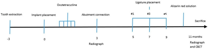
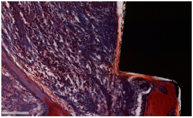
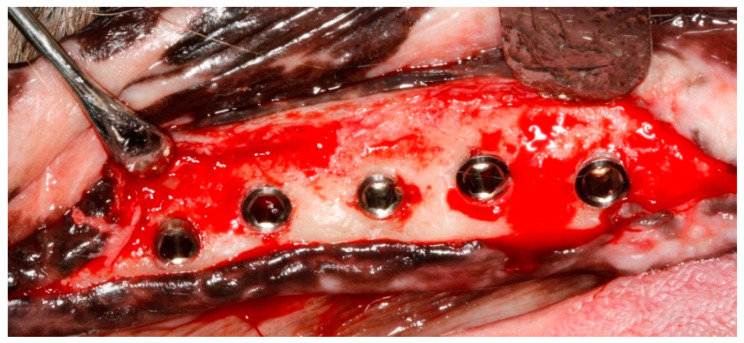
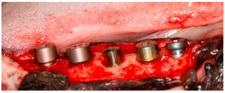
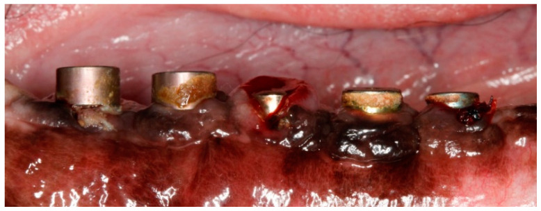
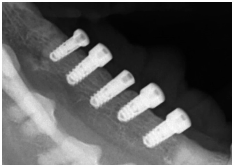
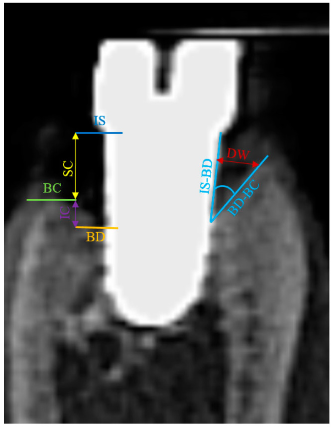
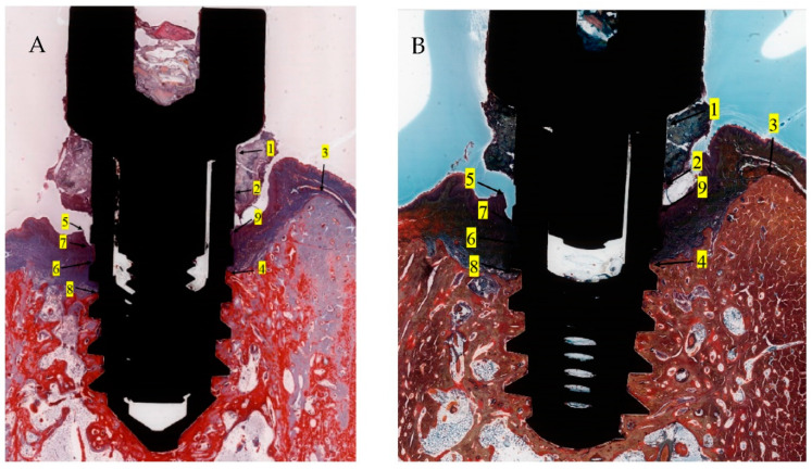
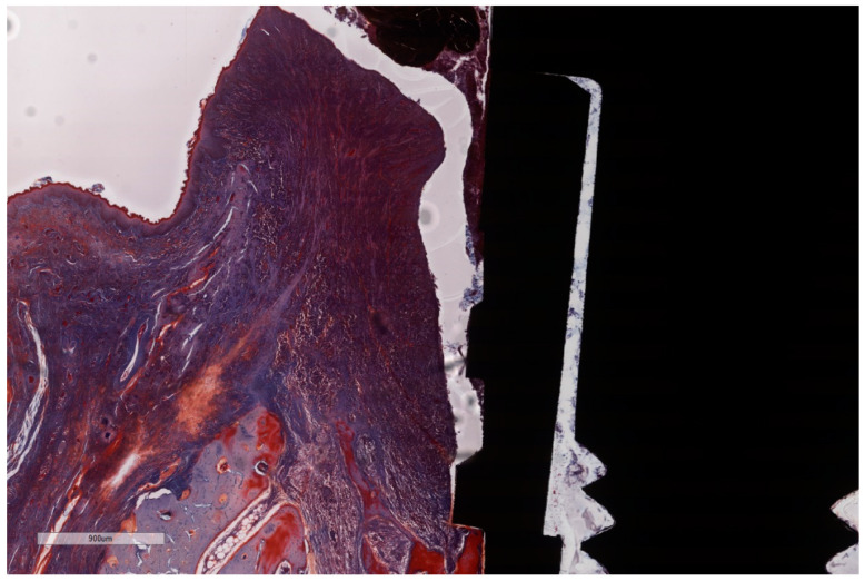
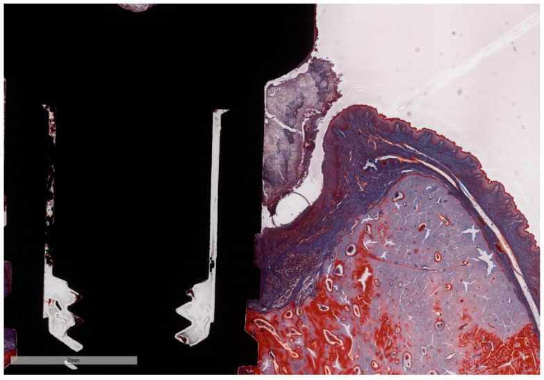
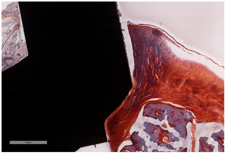
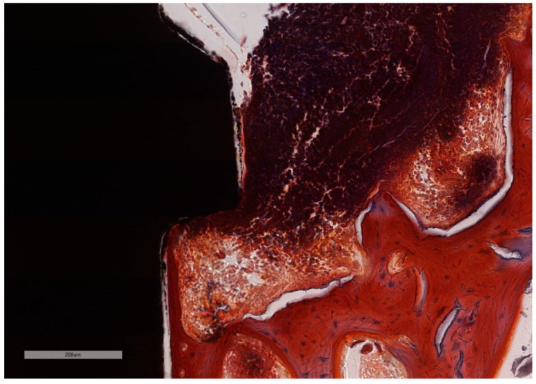
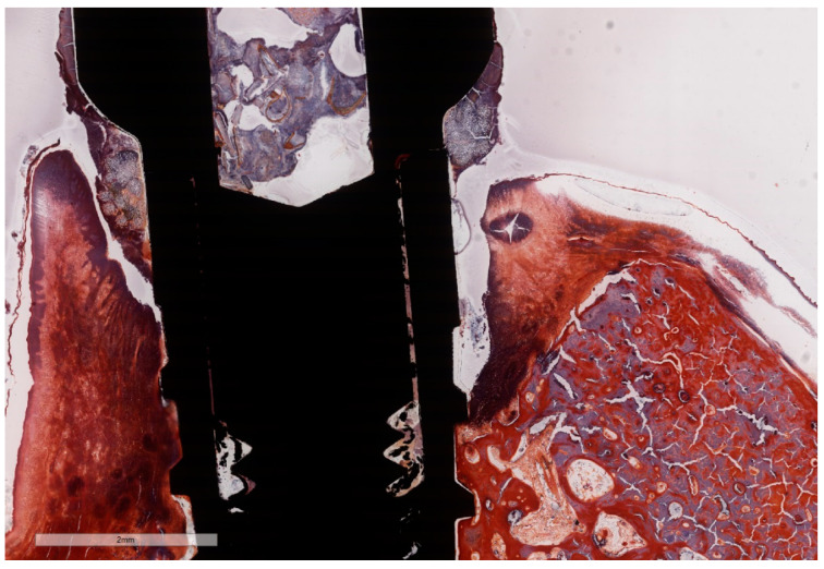
Three months after tooth extraction, five dental implants were placed in the dog hemimandible. Two months after abutment placement, ligatures were placed subsequently two months apart on alternate implants, while both intermediate implants were left without ligatures. Ligatures were kept in place during the entire experiment, and no plaque control measures were taken. Eleven months post-implantation, the animal was sacrificed. Undecalcified ground sections were cut, stained with Masson Goldner and MOVAT Pentachrome and evaluated by light microscopy. Soft and hard tissue loss was assessed using histomorphometric and CBCT parameters.
All NLI presented deep false peri-implant pockets on the oral aspect and pronounced vertical bone resorption on the buccal aspect. After 2, 4 and 6 months, during the breakdown period, more than 30% of the bone was lost in LI in all directions, while, despite immediate vicinity, NLI displayed less destruction. Intense inflammation, typical for induced peri-implantitis, was present, with similar intensity in LI as NLI, but in different parts of the lesions. Morphometry confirmed intense soft tissue inflammation, more bone resorption and higher amounts of infiltrated connective tissue in LI when compared with NLI.
Within the limits of the present pilot study, the adequacy of the experimental dog model based on ligature-induced peri-implantitis was able to be successfully challenged by non-ligature models of spontaneously occurring peri-implant inflammation, while meeting the requirements for experimental designs with a very small numbers of animals. The influence of implants with severe peri-implantitis on adjacent implants resulted in less than expected tissue loss in the latter accession numbers.
通过在同一无牙狗半侧下颌骨上交替、连续放置结扎丝,对种植体周围的软硬组织破坏情况进行组织学和放射学评估。同时评估结扎种植体(LI)对相邻未结扎种植体(NLI,作为可能自然诱发的种植体周围炎)的影响。
拔牙3个月后,在狗半侧下颌骨植入5枚牙种植体。基台植入2个月后,每隔2个月在交替的种植体上放置结扎丝,而中间的2枚种植体不放置结扎丝。在整个实验过程中结扎丝保持在位,且未采取菌斑控制措施。植入11个月后,处死动物。制备不脱钙磨片,用马松-戈德纳染色法和莫瓦特五色染色法染色,通过光学显微镜进行评估。使用组织形态计量学和锥形束计算机断层扫描(CBCT)参数评估软硬组织丧失情况。
所有NLI在口腔侧均出现较深的假性种植体周围袋,在颊侧出现明显的垂直骨吸收。在2、4和6个月的破坏期内,LI在各个方向上均有超过30%的骨丢失,而尽管距离很近,NLI的破坏程度较小。存在典型的诱导性种植体周围炎的强烈炎症,LI和NLI的炎症强度相似,但病变部位不同。形态计量学证实,与NLI相比,LI存在强烈的软组织炎症、更多的骨吸收和更高量的浸润结缔组织。
在本初步研究的范围内,基于结扎诱导的种植体周围炎的实验狗模型的适用性能够被自发发生的种植体周围炎症的非结扎模型成功挑战,同时满足使用极少量动物的实验设计要求。患有严重种植体周围炎的种植体对相邻种植体的影响导致后者的组织丧失少于预期数量。