Feng Cui, Zhou Ziling, Huang Qiuhan, Meng Xiaoyan, Li Zhen, Wang Yanchun
Departments of Radiology, Tongji Hospital, Tongji Medical College, Huazhong University of Science and Technology, Wuhan 430030, China.
Life (Basel). 2022 Sep 28;12(10):1510. doi: 10.3390/life12101510.
The aim was to evaluate the feasibility of radiomics features based on diffusion-weighted imaging (DWI) at high -values for grading bladder cancer and to compare the possible advantages of high--value DWI over the standard -value DWI.
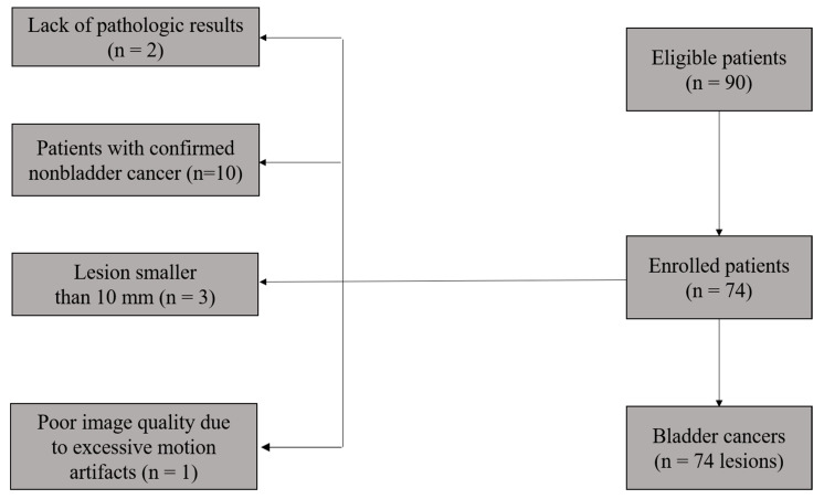
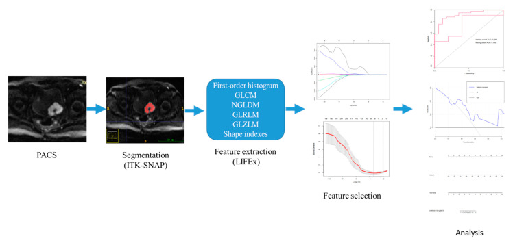
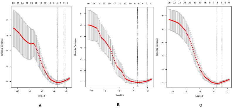
Seventy-four participants with bladder cancer were included in this study. DWI sequences using a 3 T MRI with -values of 1000, 1700, and 3000 s/mm were acquired, and the corresponding ADC maps were generated, followed with feature extraction. Patients were randomly divided into training and testing cohorts with a ratio of 8:2. The radiomics features acquired from the ADC, ADC, and ADC maps were compared between low- and high-grade bladder cancers by using the Wilcox analysis, and only the radiomics features with significant differences were selected. The least absolute shrinkage and selection operator method and a logistic regression were performed for the feature selection and establishing the radiomics model. A receiver operating characteristic (ROC) analysis was conducted to assess the diagnostic performance of the radiomics models.
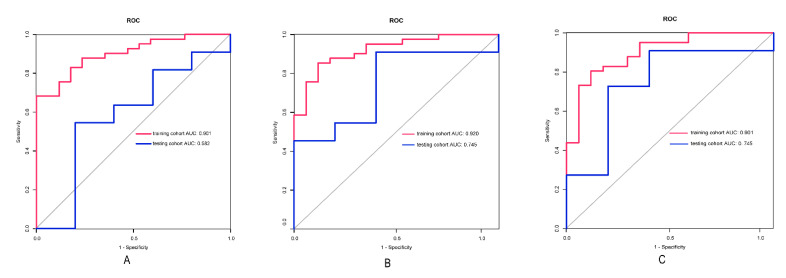
In the training cohorts, the AUCs of the ADC, ADC, and ADC model for discriminating between low- from high-grade bladder cancer were 0.901, 0.920, and 0.901, respectively. In the testing cohorts, the AUCs of ADC, ADC, and ADC were 0.582, 0.745, and 0.745, respectively.
The radiomics features extracted from the ADC maps could improve the diagnostic accuracy over those extracted from the conventional ADC maps.
目的是评估基于高b值扩散加权成像(DWI)的影像组学特征在膀胱癌分级中的可行性,并比较高b值DWI相对于标准b值DWI可能具有的优势。
本研究纳入了74例膀胱癌患者。采用3T磁共振成像获取b值为1000、1700和3000s/mm²的DWI序列,并生成相应的表观扩散系数(ADC)图,随后进行特征提取。患者按8:2的比例随机分为训练组和测试组。采用Wilcox分析比较低级别和高级别膀胱癌在ADC、ADC²和ADC³图上获取的影像组学特征,仅选择具有显著差异的影像组学特征。采用最小绝对收缩和选择算子方法及逻辑回归进行特征选择并建立影像组学模型。进行受试者工作特征(ROC)分析以评估影像组学模型的诊断性能。
在训练组中,用于区分低级别和高级别膀胱癌的ADC、ADC²和ADC³模型的曲线下面积(AUC)分别为0.901、0.920和0.901。在测试组中,ADC、ADC²和ADC³的AUC分别为0.582、0.745和0.745。
从ADC³图中提取的影像组学特征比从传统ADC图中提取的特征能提高诊断准确性。