Department of Anatomy and Medical Imaging, Faculty of Medical and Health Sciences, University of Auckland, Auckland, New Zealand.
Department of Surgery, Faculty of Medical and Health Sciences, University of Auckland, Auckland, New Zealand.
Sci Rep. 2022 Oct 28;12(1):18191. doi: 10.1038/s41598-022-23087-y.
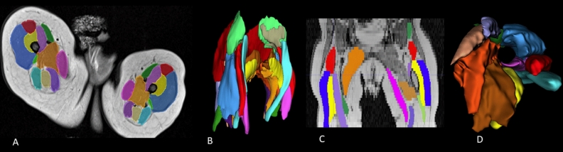
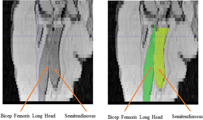
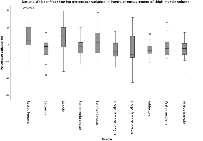
To assess intra-rater and inter-rater reliability of the manual segmentation of Magnetic Resonance Imaging (MRI) for the in vivo measurement of infant muscle volume of the knee extensor and flexor muscles by two raters. Muscles of the knee extensor and flexor muscle of ten typically developing infants (86 days ± 7 days) were scanned with MRI (Proton density sequence). Scans were then segmented using Slicer software, and volumes rendered by two raters. Intra-rater and inter-rater reliability were assessed using intra-class correlation (ICC), with mean difference (MD), standard error of the mean (SEM), and minimal detectable change (MDC) for each muscle calculated. ICCs for Intra-rater reliability of the segmentation process for the muscle volume of the muscles of the knee extensors and flexor muscles were 0.901-0.972, and 0.776-0.945 respectively, with inter-rater reliabilities between 0.914-0.954 and 0.848-0.978, for the knee extensor and flexors muscles respectively. For intra-rater reliability, MD ≤ - 0.47 cm, MDCs for were < 1.09 cm and for inter-rater MD ≤ - 1.40 cm, MDCs for were < 1.63 cm for all muscles. MRI segmentation for muscle volumes showed good to excellent reliability, though given the small volumes of the muscles themselves, variations between raters are amplified. Care should be taken in the reporting and interpretation of infant muscle volume.
评估两位评估者手动分割磁共振成像 (MRI) 以测量活体婴儿膝伸肌和屈肌肌肉体积的内部和外部可靠性。十名典型发育婴儿(86 天±7 天)的膝关节伸肌和屈肌肌肉进行 MRI(质子密度序列)扫描。然后使用 Slicer 软件对扫描进行分割,并由两位评估者渲染体积。使用组内相关系数 (ICC) 评估内部和外部可靠性,为每个肌肉计算平均差异 (MD)、均数标准差 (SEM) 和最小可检测变化 (MDC)。膝关节伸肌和屈肌肌肉体积分割过程的内部可靠性的 ICC 分别为 0.901-0.972 和 0.776-0.945,膝关节伸肌和屈肌的外部可靠性分别为 0.914-0.954 和 0.848-0.978。对于内部可靠性,MD≤-0.47cm,MDCs 为<1.09cm,对于外部可靠性,MD≤-1.40cm,MDCs 为<1.63cm。MRI 分割肌肉体积的可靠性较好,但由于肌肉本身的体积较小,评估者之间的差异被放大。在报告和解释婴儿肌肉体积时应谨慎。