Department of Orthopaedics and Sports Orthopaedics, Klinikum rechts der Isar, Technical University of Munich, Ismaninger Str. 25, 81675, Munich, Germany.
Institute for AI and Informatics in Medicine, Technical University of Munich, Munich, Germany.
Eur Radiol. 2023 Mar;33(3):1537-1544. doi: 10.1007/s00330-022-09184-6. Epub 2022 Oct 29.
To develop a two-phased deep learning sorting algorithm for post-X-ray image acquisition in order to facilitate large musculoskeletal image datasets according to their anatomical entity.
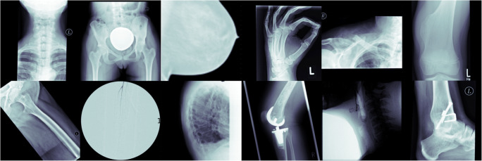
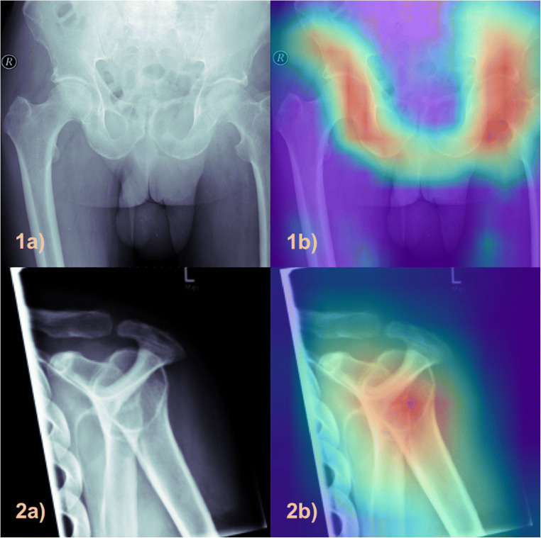
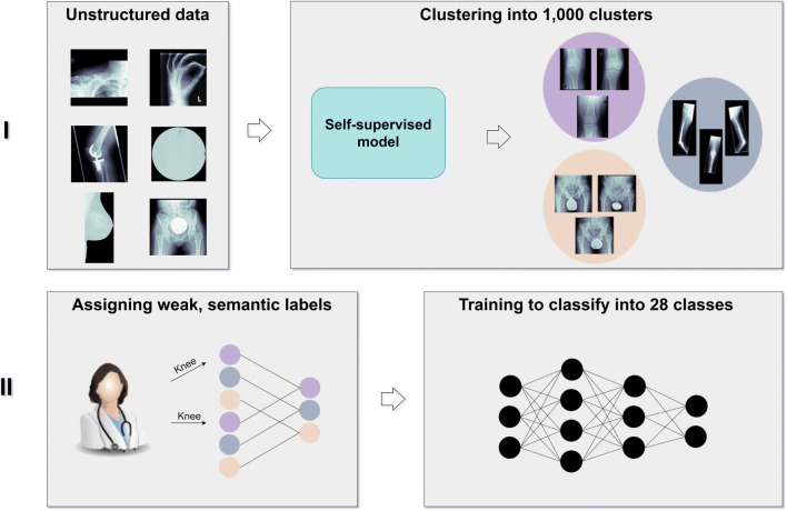
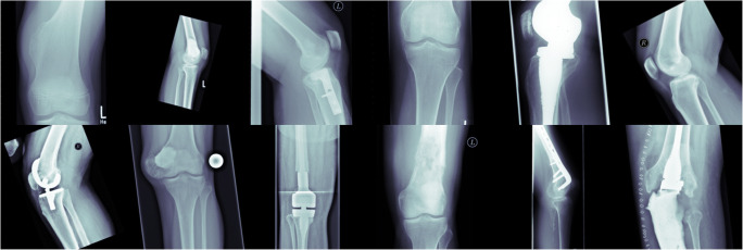
In total, 42,608 unstructured and pseudonymized radiographs were retrieved from the PACS of a musculoskeletal tumor center. In phase 1, imaging data were sorted into 1000 clusters by a self-supervised model. A human-in-the-loop radiologist assigned weak, semantic labels to all clusters and clusters with the same label were merged. Three hundred thirty-two non-musculoskeletal clusters were discarded. In phase 2, the initial model was modified by "injecting" the identified labels into the self-supervised model to train a classifier. To provide statistical significance, data split and cross-validation were applied. The hold-out test set consisted of 50% external data. To gain insight into the model's predictions, Grad-CAMs were calculated.
The self-supervised clustering resulted in a high normalized mutual information of 0.930. The expert radiologist identified 28 musculoskeletal clusters. The modified model achieved a classification accuracy of 96.2% and 96.6% for validation and hold-out test data for predicting the top class, respectively. When considering the top two predicted class labels, an accuracy of 99.7% and 99.6% was accomplished. Grad-CAMs as well as final cluster results underlined the robustness of the proposed method by showing that it focused on similar image regions a human would have considered for categorizing images.
For efficient dataset building, we propose an accurate deep learning sorting algorithm for classifying radiographs according to their anatomical entity in the assessment of musculoskeletal diseases.
• Classification of large radiograph datasets according to their anatomical entity. • Paramount importance of structuring vast amounts of retrospective data for modern deep learning applications. • Optimization of the radiological workflow and increase in efficiency as well as decrease of time-consuming tasks for radiologists through deep learning.
开发一种两阶段深度学习排序算法,以便根据解剖实体对 X 射线后获取的图像进行分类,从而方便处理大型肌肉骨骼影像数据集。
共从肌肉骨骼肿瘤中心的 PACS 中检索了 42608 张未结构化和匿名化的射线照片。在第一阶段,通过自监督模型将影像学数据分为 1000 个聚类。一位人类专家放射科医生对所有聚类赋予了弱语义标签,并将具有相同标签的聚类合并。丢弃了 332 个非肌肉骨骼聚类。在第二阶段,通过将识别出的标签“注入”到自监督模型中,对初始模型进行了修改,以训练分类器。为了提供统计意义,应用了数据分割和交叉验证。保留测试集由 50%的外部数据组成。为了深入了解模型的预测,计算了 Grad-CAMs。
自监督聚类得到了归一化互信息为 0.930 的较高分数。专家放射科医生识别出 28 个肌肉骨骼聚类。经过修改的模型在验证和保留测试数据中对预测最高类别的准确率分别为 96.2%和 96.6%。当考虑预测的前两个类标签时,准确率达到了 99.7%和 99.6%。Grad-CAMs 以及最终的聚类结果强调了该方法的稳健性,表明它专注于人类在对图像进行分类时会考虑的类似图像区域。
为了实现高效的数据构建,我们提出了一种准确的深度学习排序算法,用于根据肌肉骨骼疾病评估中图像的解剖实体对射线照片进行分类。
• 根据解剖实体对大型射线照片数据集进行分类。
• 对于现代深度学习应用,构建大量回顾性数据的重要性。
• 通过深度学习优化放射科工作流程,提高效率,减少放射科医生的耗时任务。