Department of Orthopaedics and Sports Orthopaedics, Klinikum rechts der Isar, Technical University of Munich, Munich, Germany.
Institute for AI and Informatics in Medicine, Technical University of Munich, Munich, Germany.
Eur Radiol. 2024 Oct;34(10):6629-6638. doi: 10.1007/s00330-024-10672-0. Epub 2024 Mar 15.
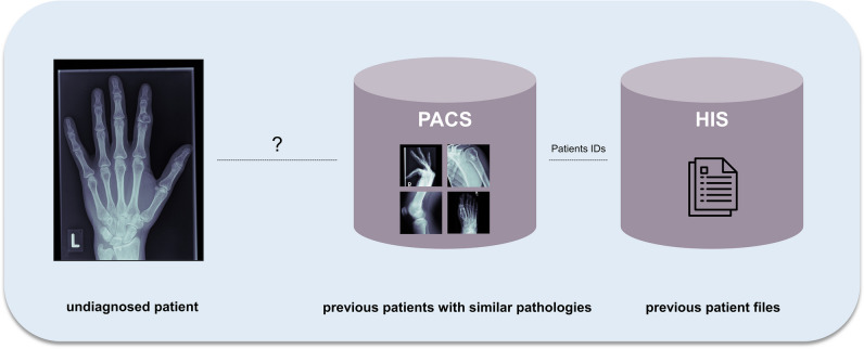
To develop an algorithm to link undiagnosed patients to previous patient histories based on radiographs, and simultaneous classification of multiple bone tumours to enable early and specific diagnosis.
For this retrospective study, data from 2000 to 2021 were curated from our database by two orthopaedic surgeons, a radiologist and a data scientist. Patients with complete clinical and pre-therapy radiographic data were eligible. To ensure feasibility, the ten most frequent primary tumour entities, confirmed histologically or by tumour board decision, were included. We implemented a ResNet and transformer model to establish baseline results. Our method extracts image features using deep learning and then clusters the k most similar images to the target image using a hash-based nearest-neighbour recommender approach that performs simultaneous classification by majority voting. The results were evaluated with precision-at-k, accuracy, precision and recall. Discrete parameters were described by incidence and percentage ratios. For continuous parameters, based on a normality test, respective statistical measures were calculated.
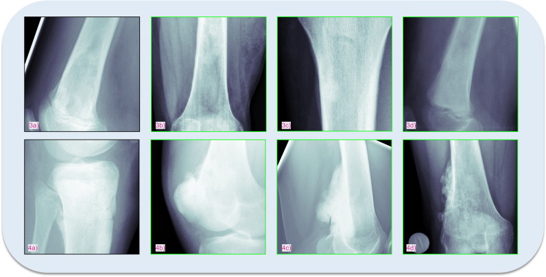
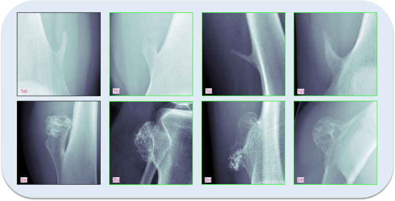
Included were data from 809 patients (1792 radiographs; mean age 33.73 ± 18.65, range 3-89 years; 443 men), with Osteochondroma (28.31%) and Ewing sarcoma (1.11%) as the most and least common entities, respectively. The dataset was split into training (80%) and test subsets (20%). For k = 3, our model achieved the highest mean accuracy, precision and recall (92.86%, 92.86% and 34.08%), significantly outperforming state-of-the-art models (54.10%, 55.57%, 19.85% and 62.80%, 61.33%, 23.05%).
Our novel approach surpasses current models in tumour classification and links to past patient data, leveraging expert insights.
The proposed algorithm could serve as a vital support tool for clinicians and general practitioners with limited experience in bone tumour classification by identifying similar cases and classifying bone tumour entities.
• Addressed accurate bone tumour classification using radiographic features. • Model achieved 92.86%, 92.86% and 34.08% mean accuracy, precision and recall, respectively, significantly surpassing state-of-the-art models. • Enhanced diagnosis by integrating prior expert patient assessments.
开发一种基于 X 光片将未确诊患者与既往病史联系起来的算法,并同时对多种骨肿瘤进行分类,以实现早期和特异性诊断。
本回顾性研究对 2000 年至 2021 年期间我们数据库中的数据进行了 curation,由两名骨科医生、一名放射科医生和一名数据科学家进行 curation。符合条件的患者需要具备完整的临床和治疗前放射学数据。为确保可行性,纳入了十种最常见的经组织学证实或经肿瘤委员会决定的原发性肿瘤实体。我们实施了 ResNet 和变压器模型以建立基线结果。我们的方法使用深度学习提取图像特征,然后使用基于哈希的最近邻推荐方法对 k 个与目标图像最相似的图像进行聚类,该方法通过多数投票进行同时分类。使用精度@k、准确性、精度和召回率来评估结果。离散参数用发生率和百分比比值来描述。对于连续参数,根据正态性检验,计算相应的统计量。
纳入了 809 名患者(1792 张 X 光片;平均年龄 33.73±18.65 岁,范围 3-89 岁;443 名男性),其中 Osteochondroma(骨软骨瘤)(28.31%)和 Ewing sarcoma(尤文肉瘤)(1.11%)分别是最常见和最不常见的实体。数据集分为训练集(80%)和测试集(20%)。对于 k=3,我们的模型达到了最高的平均准确率、精度和召回率(92.86%、92.86%和 34.08%),明显优于最先进的模型(54.10%、55.57%、19.85%和 62.80%、61.33%、23.05%)。
我们的新方法在肿瘤分类和与既往患者数据的链接方面优于现有模型,利用了专家的见解。
该算法可以作为一种重要的支持工具,为经验有限的骨科医生和全科医生提供帮助,通过识别相似病例和分类骨肿瘤实体来协助诊断。
利用放射学特征实现了准确的骨肿瘤分类。
模型达到了 92.86%、92.86%和 34.08%的平均准确率、精度和召回率,明显优于最先进的模型。
通过整合以前的专家患者评估来增强诊断。